
Presentation
Right upper quadrant pain.
Patient Data
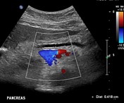
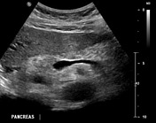
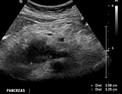
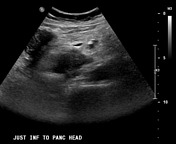
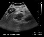
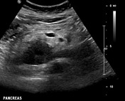
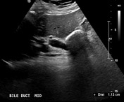
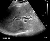
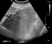
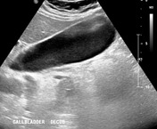

Selected ultrasound images showing a hypoechogenic solid mass at the pancreatic head/uncinate process leading to intra and extrahepatic biliary tree dilatation.


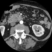
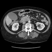
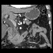
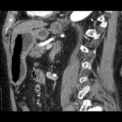
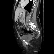
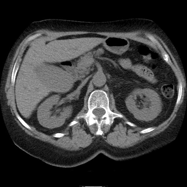
There is a mass in the uncinate process of the pancreas centered inferior, posterior, and medial to the ampulla. As a result, there is only mild prominence of the main pancreatic duct. There is a somewhat elongated low density structure within the tumor, which either represents a focally dilated side branch ducts or else central necrotic/cystic change. There is intra-hepatic duct dilatation and the common bile duct measures 9 mm in diameter. Complete loss of fat planes between the tumor and segment deep three of the duodenum.
Normal variant anatomy with the left and right hepatic artery arising off the celiac axis. Celiac axis, hepatic arteries, superior mesenteric artery are clear of tumor.
The portal vein is clear of tumor. There is a short segment contact of abnormal soft tissue at the level of the tumor with a left tributary to the superior mesenteric vein. The main superior mesenteric vein is clear of tumor.
There are no abnormal lymph nodes by size or appearance in the chest, abdomen or pelvis.
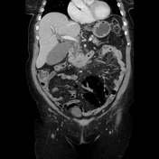
Several subcentimeter low density liver lesions are seen, best appreciated on coronal portal venous phase portal venous phase. These are indeterminate but should be regarded with suspicion for metastasis.
A number of simple renal cysts are noted, otherwise the kidneys are normal. The adrenal glands and spleen are normal. No concerning bone lesion.

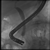
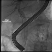

ERCP was performed for distal CBD stenting.
Case Discussion
This case illustrates an uncinate process of pancreas carcinoma. As shown on dedicated arterial and portal venous phases, the regional relevant vasculature is clear of tumor, but liver metastasis are already present, which already makes it a stage IV (M1 any T any N).
EUS FNA has been performed to the mass and one of the liver metastasis:
Microscopy: Slides show a population of malignant tumor cells forming variably sized cohesive clusters. These cells have pleomorphic, hyperchromatic nuclei with prominent nucleoli and there is a moderate amount of cytoplasm. The findings are consistent with adenocarcinoma.
Macroscopy: Endoscopic ultrasound-guided FNA of pancreatic uncinate mass. Multiple passes 1 air-dried and 1 alcohol-fixed smears prepared. Cell block prepared. Cytologist in attendance.
Conclusion:
- pancreatic uncinate mass, EUS-guided FNA: Positive for adenocarcinoma
- liver mass, EUS-guided FNA: Positive for metastatic adenocarcinoma


 Unable to process the form. Check for errors and try again.