
Presentation
28/40 pregnant. Right upper quadrant pain and fever. No abnormality detected on ultrasound.
Patient Data


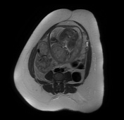
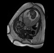

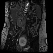
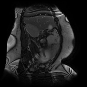
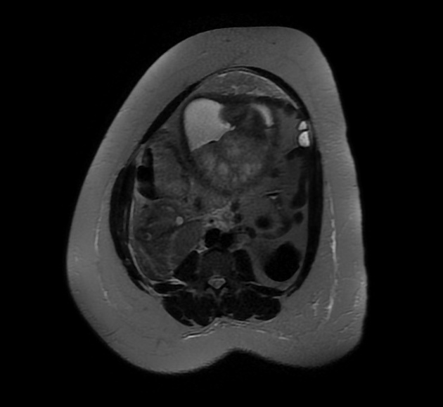
Intrauterine pregnancy confirmed but not interrogated in detail. The gravid uterus displaces the cecal pole and appendix out of the right iliac fossa, and the cecal pole is tilted posteriorly. An acute inflammatory mass is present posteriorly, with edema of the fat and free fluid throughout the right side of the abdomen. Appendicoliths are seen within the distended edematous appendix. The appearances are consistent with acute appendicitis.
Histopathology
Macroscopic: Appendix covered in fibrinopurulent exudate 60x14mm.
Microscopic: This appendix shows abundant transmural acute inflammation and extensive tissue necrosis. There is no evidence of dysplasia or malignancy.
Conclusion: Appendix - acute gangrenous appendicitis
Case Discussion
The MRI findings are consistent with acute appendicitis with an inflammatory mass in the right side of the abdomen, and the findings were confirmed at surgery. Due to a change in the position of the appendix during pregnancy, localization of pain varies. The position as seen here also made an ultrasound diagnosis difficult to make. MRI, where available, is recommended as the second line modality by the ACR Appropriateness Criteria 1,2.


 Unable to process the form. Check for errors and try again.