
Presentation
Intravenous drug user, septic.
Patient Data
Age: 35 years
Gender: Female


Download
Info

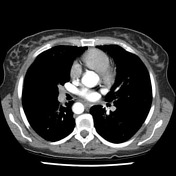
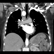
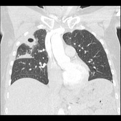
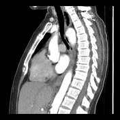
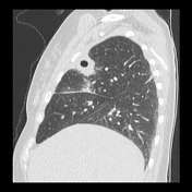
In the right upper lobe there are two peripheral and somewhat wedge shaped areas of consolidation with internal cavitations that are surrounded by a thick enhancing rim. The lungs and pleural spaces are otherwise unremarkable.
Case Discussion
Chest radiograph from a previous admission 3 months ago was normal (not shown). In this clinical settings, the imaging appearances are those of lung abscesses, likely due to heart or peripheral source of seeding. Echo-cardiogram has been recommended with view to check for right heart valve vegetations.


 Unable to process the form. Check for errors and try again.