
Presentation
Progressive left shoulder pain, swelling, and restricted range of movements.
Patient Data
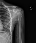
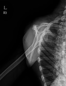
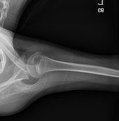

There is aggressive (moth-eaten) osteolytic lesion involving the left scapula, mainly its spine.
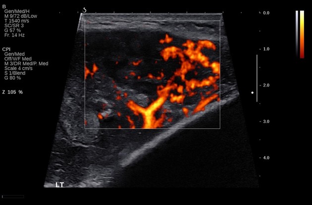
Well defined heterogeneous lesion (6.8 X 10.5 cm) at left shoulder mainly involves the left scapula, left supraspinatus muscle, and left infraspinatus muscle. It shows increased vascularity on Doppler images. Indistinctness and irregularity of hyperechoic cortex is suspicious for bony erosion of the left scapula.


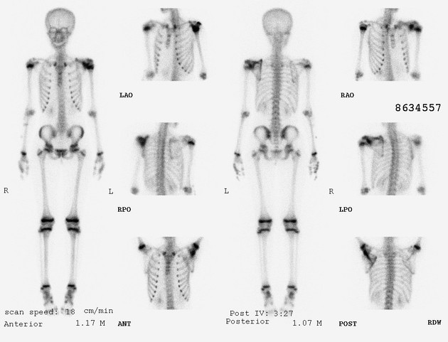
Whole body bone scan: there is increased radiotracer uptake in the left scapula, suspicious for malignancy. No other significant abnormal bone uptake to suggest skeletal metastasis.
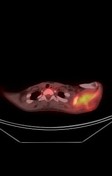
PET-CT whole body scan: there is increased FDG uptake in the soft tissue mass involving left scapula, suggestive of malignancy. No other significant abnormal FDG uptake to suggest distant metastasis.
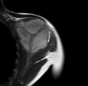

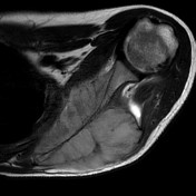
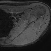
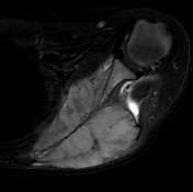
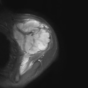
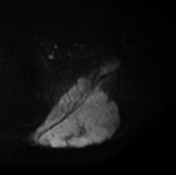
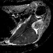
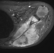
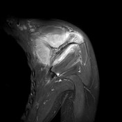
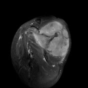

Extensive soft tissue mass (9.5 X 6 X 7.6 cm) involving left scapula (including acromion and coracoid) with invasion of the supraspinatus, infraspinatus, and subscapularis muscles.
This mass shows diffuse high signal with low signal striation on T2 WI, prominent enhancement, and diffusion restriction.
No definite osteoid matrix/calcification in soft tissue component.
Multiple borderline lymph nodes in left supraclavicular fossa and axillary area.
Edematous changes at supraspinatus, infraspinatus, and teres minor muscles.
Case Discussion
Tissue biopsy was taken from the mass and Ewing's sarcoma was confirmed.


 Unable to process the form. Check for errors and try again.