
Patient Data
Note: This case has been tagged as "legacy" as it no longer meets image preparation and/or other case publication guidelines.

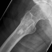
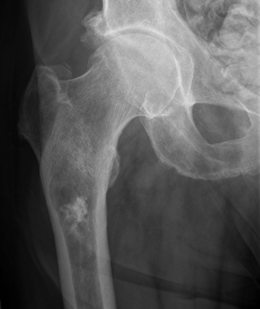
An intramedullary lesion is noted with associated endosteal scalloping and chondral calcification. No periosteal reaction or extra-osseous mass is evident.
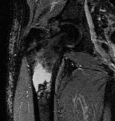
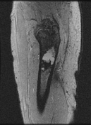

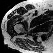

Selected images from an MRI demonstrate the intramedullary lesion to have low T1 and high T2 signal. Endosteal scalloping is present laterally but there is no extra-osseous extension.
Features are consistent with a low-grade chondroid lesion, likely a chondrosarcoma.
Case Discussion
The patient went on to have surgery.
Histology
Gross description: The specimen showed a lobulated poorly demarcated blue-tinged intramedullary lesion that measures 3.5 x 2.8 x 2.5 cm. The lesion is completely confined within the medullary cavity with areas of endosteal scalloping but there is no breakthrough the cortical bone as well as no soft tissue extension. Negative for dedifferentiation
Final diagnosis: chondrosarcoma (intermediate grade, grade 2/3)


 Unable to process the form. Check for errors and try again.