
Presentation
Sepsis, intravenous drug user.
Patient Data
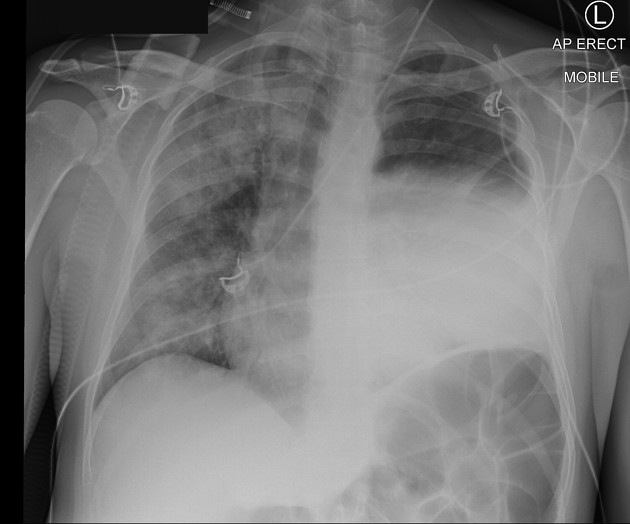
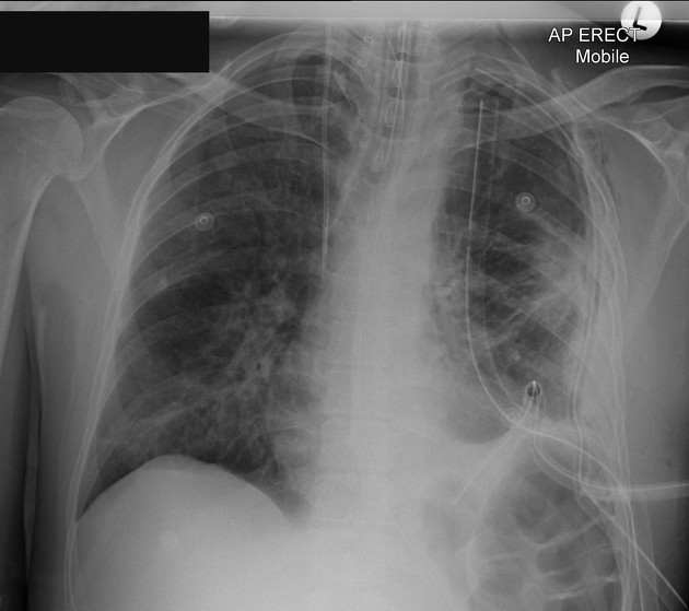
Confluent left lung consolidation. Peripheral air-fluid level in the left suggestive of an empyema. There is a mass effect with rightward mediastinal deviation, even allowing for mildly rotated radiograph. Confluent right lung consolidation.


ETT in situ. NGT with tip and side-holes in the stomach.
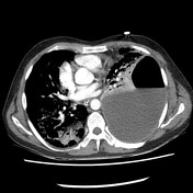
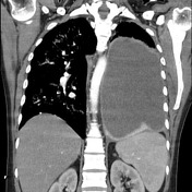
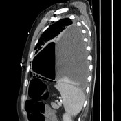
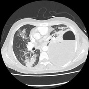
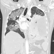
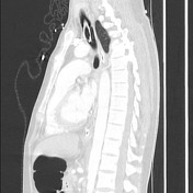
Multifocal, patchy bilateral lobar consolidation with air bronchograms are most in keeping with bacterial pneumonia. Large loculated left-sided pleural effusion with an air-fluid level and pleural thickening is consistent with empyema. There is associated compressive atelectasis of the left lung and mild mediastinal shift. Heart and great vessels are within normal limits. Non-pathologically enlarged mediastinal lymph nodes are likely reactive. No suspicious osseous lesion is identified.
Left empyema drained; three large-bore left pleural ICC. Re-expansion of the left lung with persistent left base atelectasis,small amount of pleural fluid, as well as patchy airspace opacities in the midzone that are likely related to re-expansion. Improved right lung patchy airspace opacities. Right-sided central venous line and ETT are in situ.
Case Discussion
Left thoracotomy has been performed with drainage of frank pus.


 Unable to process the form. Check for errors and try again.