
Presentation
Severe right pleuritic pain. No clinical evidence of infection.
Patient Data
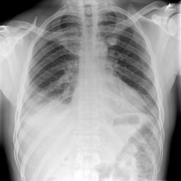
Poor inspiratory effort. Mild rotation of the patient to the left.
Patchy consolidation obscuring the right hemidiaphragm consistent with right lower lobe pathology. Left lung is clear.
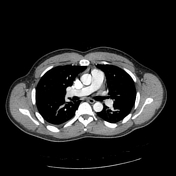


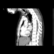


Low density filling defects representing acute PE in the anterior segmental branch of the right lower lobe pulmonary artery and subsegmental branches of the middle and left lower lobe pulmonary arteries. No pulmonary artery enlargement.
Wedge-shaped ground-glass opacity of the anterior segment of the right lower lobe consistent with pulmonary infarction; several internal gas filled foci ? cavitation. Subsegmental atelectasis within the middle lobe and posterior lower lobe.
Very shallow right-sided pleural effusion. No pericardial effusion.
Incidental note of a 1.5 cm hypervascular hepatic lesion of central segment 2, differential including focal nodular hyperplasia and flash-filling hemangioma, neither diagnosis is usually of any significance. Several low density liver lesions, too small to characterize, most likely cysts. No other findings of concern of the partially imaged upper abdomen.
Conclusion:
1. Bilateral segmental PEs
2. Pulmonary infarction (with possible cavitation) of the anterior segment of the right lower lobe. No evidence of pneumonia.
Case Discussion
Bilateral acute PEs of the segmental pulmonary arteries with a right lower lobe pulmonary infarction.


 Unable to process the form. Check for errors and try again.