
Patient Data
Gender: Unknown
Download
Info

The labeled structures are (excluding the correct side):
- humeral head
- subscapularis muscle
- glenohumeral joint
- inferior angle of the scapula
- infraspinatus muscle
- spine of the scapula
- axillary artery
- axillary vein
- trapezius muscle
- pectoralis major muscle
- pectoralis minor muscle
- supraspinatus muscle
- subclavian vein
- subclavian artery
- subclavius muscle
- breast
- rhomboid major muscle
- costal cartilage
- internal thoracic (mammary) artery
- rhomboid minor muscle
- epicardial fat pad
- right upper lobe bronchus
- bronchus intermedius
- right subclavian artery
- medial end of the clavicle
- right interlobar pulmonary artery
- sternoclavicular joint
- right upper lobar pulmonary artery
- right superior pulmonary vein
- internal jugular vein
- right inferior pulmonary vein
- right brachiocephalic vein
- arch of the azygos vein
- body of the sternum
- right atrium
- common carotid artery
- right main bronchus
- superior vena cava
- sternomanubrial joint
- sternoxiphoid joint
- xiphoid process of the sternum
- brachiocephalic trunk
- inferior vena cava
- suprasternal (jugular) notch
- left atrium
- isthmus of the thyroid gland
- trachea
- posterior intercostal artery
- left brachiocephalic vein
- right pulmonary artery
- manubrium of the sternum
- right atrial appendage (auricle)
- ascending aorta
- esophagus
- left lobe of the thyroid gland
- left main bronchus
- descending aorta
- left common carotid artery
- posterior intercostal artery
- vertebral artery
- left subclavian artery
- aortic isthmus
- aortic arch
- right ventricle
- interventricular septum
- pulmonary trunk
- left superior pulmonary vein
- left inferior pulmonary vein
- left atrial appendage (auricle)
- left pulmonary artery
- left lower lobar bronchus
- right ventricle
- left upper lobar bronchus
- left interlobar pulmonary artery
- left upper lobar pulmonary artery
- left ventricle
- latissimus dorsi muscle
- serratus anterior muscle
Download
Info
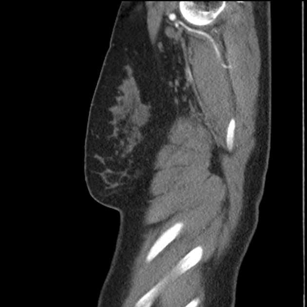
The same normal abdominal CT arterial phase without labels for reference.
Case Discussion
The labeled structures are (excluding the correct side):
- humeral head
- subscapularis muscle
- glenohumeral joint
- inferior angle of the scapula
- infraspinatus muscle
- spine of the scapula
- axillary artery
- axillary vein
- trapezius muscle
- pectoralis major muscle
- pectoralis minor muscle
- supraspinatus muscle
- subclavian vein
- subclavian artery
- subclavius muscle
- breast
- rhomboid major muscle
- costal cartilage
- internal thoracic (mammary) artery
- rhomboid minor muscle
- epicardial fat pad
- right upper lobe bronchus
- bronchus intermedius
- right subclavian artery
- medial end of the clavicle
- right interlobar pulmonary artery
- sternoclavicular joint
- right upper lobar pulmonary artery
- right superior pulmonary vein
- internal jugular vein
- right inferior pulmonary vein
- right brachiocephalic vein
- arch of the azygos vein
- body of the sternum
- right atrium
- common carotid artery
- right main bronchus
- superior vena cava
- sternomanubrial joint
- sternoxiphoid joint
- xiphoid process of the sternum
- brachiocephalic trunk
- inferior vena cava
- suprasternal (jugular) notch
- left atrium
- isthmus of the thyroid gland
- trachea
- posterior intercostal artery
- left brachiocephalic vein
- right pulmonary artery
- manubrium of the sternum
- right atrial appendage (auricle)
- ascending aorta
- esophagus
- left lobe of the thyroid gland
- left main bronchus
- descending aorta
- left common carotid artery
- posterior intercostal artery
- vertebral artery
- left subclavian artery
- aortic isthmus
- aortic arch
- right ventricle
- interventricular septum
- pulmonary trunk
- left superior pulmonary vein
- left inferior pulmonary vein
- left atrial appendage (auricle)
- left pulmonary artery
- left lower lobar bronchus
- right ventricle
- left upper lobar bronchus
- left interlobar pulmonary artery
- left upper lobar pulmonary artery
- left ventricle
- latissimus dorsi muscle
- serratus anterior muscle


 Unable to process the form. Check for errors and try again.