
Presentation
Abdominal pain and distention.
Patient Data
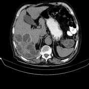


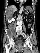
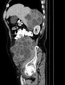

A large thick walled multiloculated cystic lesion, 146×110×80mm in diameter in the right lobe of the liver. There are also several large cystic lesions with similar appearance at right abdominopelvic spaces as well as pre-splenic region. Small foci of calcifications are noted in some lesions.
A 30 mm anterior abdominal wall defect with herniation of some omental fat are observed.
The prostate gland is enlarged.
Degenerative changes as osteophytosis are seen at the lumbar spine.
Case Discussion
Hepatic, intra- and retro-peritoneal multiloculated cystic lesions compatible with hydatid cysts. Retroperitoneal hydatid cysts are generally seen secondary to intraperitoneal (mainly hepatic) infection. Isolated occurrence is rare.


 Unable to process the form. Check for errors and try again.