
Presentation
Left shoulder pain for 3 years.
Patient Data
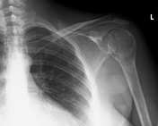
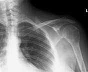
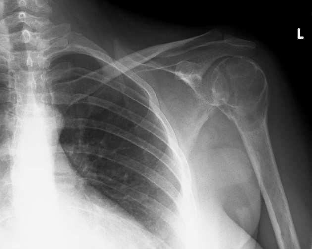
Well-defined osteolytic lesion involving the left humeral head and neck. There is a sharp demarcation line from the healthy bone. The lesion erodes the endosteum and invades the joint medially. No periosteal reaction detected. Pathological fracture through the distal third of the lesion.
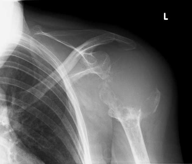
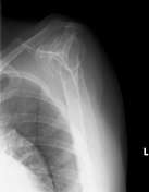
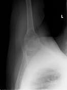
After having been left undiagnosed - and therefore, untreated - for almost 3 years, the lesion has expanded considerably, with nearly complete destruction of the proximal third of the bone and evidence of soft-tissue mass.


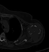
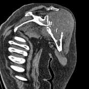
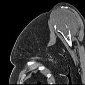
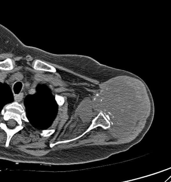
Large, heterogeneous, relatively low-density mass lesion in the humeral head with foci of calcification, mostly at its periphery. The lesion breaks through the bone circumferentially, involves the glenohumeral joint, and displaces the nearby muscles with loss of the intervening fat plane. Its low density is suggestive of a chondroid matrix.
There is a non-enlarged, rounded, axillary lymph node lacking a fatty hilum - cannot be ruled out as metastatic.
Case Discussion
Based on its radiological appearance and relatively slow growth, the lesion is most compatible with clear cell chondrosarcoma.


 Unable to process the form. Check for errors and try again.