
Presentation
24/40 pregnant. Right-sided abdominal pain and fever.
Patient Data
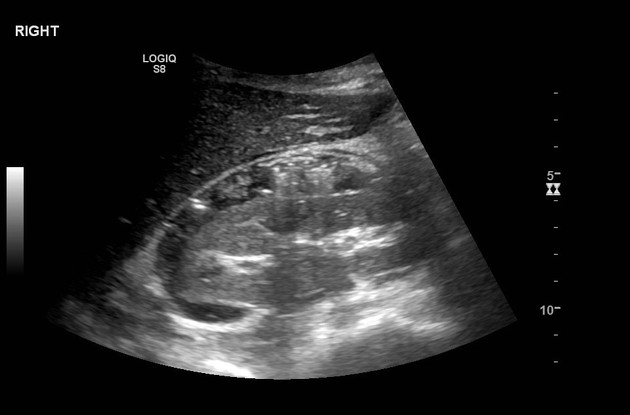
Appendix not clearly identified in right lower quadrant. Liver and gall bladder appear normal. Right kidney appears normal with no mass or hydronephrosis, but there is abnormal echogenicity in the perinephric space, with a speckled appearance. Further evaluation with MRI is recommended.
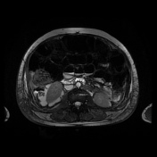

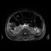
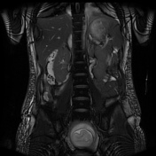

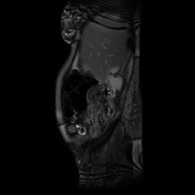
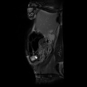

Intrauterine pregnancy confirmed but not interrogated in detail. There is a small volume of free fluid throughout the abdomen, as well as abdominal wall edema. The appendix, lying posterior to the cecum and proximal ascending colon, is abnormal in appearance. It is dilated, thick-walled and edematous. It contains appendicoliths, and the tip shows discontinuity of the wall. As a result of perforation from appendicitis, there is admixed gas and fluid in the right perinephric space. This explains the appearances on the prior ultrasound.
Histopathology
Clinical History: Appendicitis
Macroscopic: Appendix measuring 52 x 20 x 12 mm covered with fibrinous exudate.
Microscopic: Microscopy shows active transmural inflammation with perforation and peritonitis. There is no dysplasia, no evidence of malignancy and no parasites are seen.
Conclusion: Appendix - acute gangrenous appendicitis with peritonitis
Case Discussion
The ultrasound findings indicate bubbles of gas within the perinephric space - a highly unusual finding, particularly given that the kidney itself appears normal. The MRI helps to diagnose perforated acute appendicitis as the cause of the ultrasound findings and the clinical presentation. The appendix is dilated, thick-walled and contains appendicoliths. Examination in three planes enables the path of the appendix to be followed.


 Unable to process the form. Check for errors and try again.