
Presentation
Shortness of breath for one month.
Patient Data

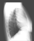
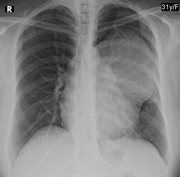
A well-defined large mediastinal mass originates from the middle mediastinum and extends to the left middle zone.
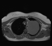

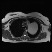
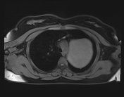
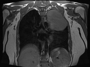
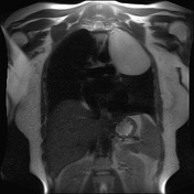
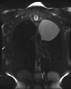
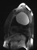

There is a well-circumscribed spherical mass lesion in the left upper zone, originating from the left aspect of the middle mediastinum, abutting the aortic arch and left main vessels without evidence of invasion into the mediastinal structures. The lesion measures ~9.5 cm in diameter. It shows high signal on T1 and T2 suggesting protein content.
Case Discussion
Bronchogenic cysts are congenital malformations of the bronchial tree They can present as a mediastinal mass that may enlarge and cause local compression. It is considered the commonest of foregut duplication cysts.
In many instances, bronchogenic cysts are asymptomatic and are found incidentally when the chest is imaged. When large, mass effect may result in bronchial obstruction leading to air trapping and respiratory distress.


 Unable to process the form. Check for errors and try again.