
Presentation
Incidental finding post MVA.
Patient Data
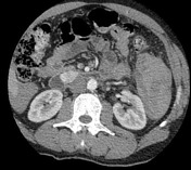


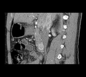
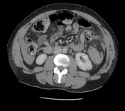

There is a 3 cm hypervascular lesion involving the D2/D3 duodenal wall. Although closely related to the uncinate process of the pancreas, it appears to have its origin in the duodenum. No necrosis or calcification identified.
Hemoperitoneum related to splenic rupture that was treated with splenic artery embolization. The spleen is enlarged.
The remainder of the study is unremarkable.
A biopsy was performed via gastroscopy and revealed:
Microscopy: The sections show strips of duodenal mucosa overlying a proliferation of spindled cells with elongate, blunt-ended, bland appearing nuclei and tapered eosinophilic cytoplasm. A rare mitotic figure is identified. Lesional cells immunoreactive with antibodies against CD117 (c-kit) and SMA and are negative with antibodies against desmin. Ki67 proliferative index is < 3%.
Conclusion: Subepithelial D2 lesion biopsy: Gastrointestinal stromal tumor (GIST).
Case Discussion
Duodenal GIST in close relation to the pancreatic head.


 Unable to process the form. Check for errors and try again.