
Presentation
Lumbar laminectomy and discectomy 1/52 ago. Since then ongoing headache and photophobia.
Patient Data


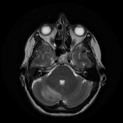
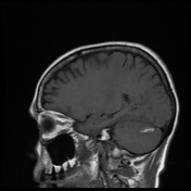
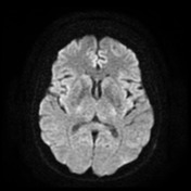
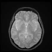
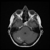
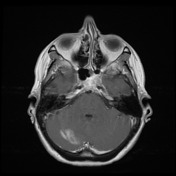

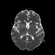
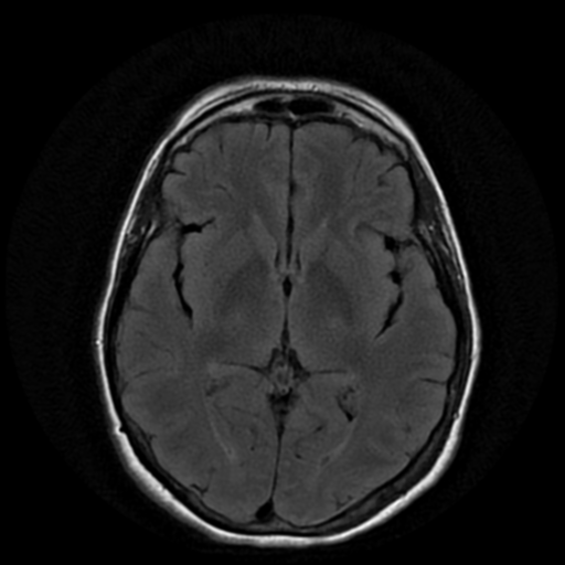
In the right cerebellar hemisphere is a parenchymal hemorrhage with thin surrounding enhancement and a small amount of edema. No nodular contrast enhancement here or elsewhere to suggest underlying lesion. No abnormal vessels on MRA. The remainder of the brain is unremarkable in appearance.
Conclusion: In this clinical setting, features are almost certainly those of a postoperative remote cerebellar hemorrhage.
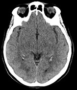
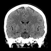
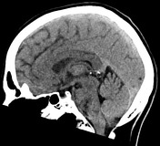
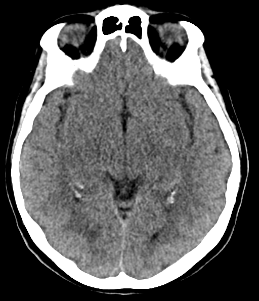
Poorly defined ovoid focus of hypoattenuation in the superior aspect of the right cerebellar hemisphere corresponds to the location of blood products demonstrated on the prior MRI. No CT evidence of recent acute hemorrhage. The remainder of the brain parenchyma is unremarkable. Ventricles and cortical sulci within normal limits. No hydrocephalus. Imaged para nasal sinuses and mastoid air cells were aerated. No osseous lesion.
Case Discussion
Remote cerebellar hemorrhages are uncommon and are important to recognize.


 Unable to process the form. Check for errors and try again.