
Remote cerebellar hemorrhage is a relatively benign complication of supratentorial craniotomy, spinal surgery, lumbar puncture and insertion of a lumboperitoneal shunt 1,2,9. It is called "remote" as the cerebellar hemorrhage is far from the location of the surgery.
On this page:
Epidemiology
A rare complication seen in 0.04% to 0.8% post craniotomy and post spinal surgeries possibly due to break in dura 8.
Clinical presentation
Most patients are asymptomatic 2. When symptomatic, delayed awakening from anesthesia and reduced level of consciousness are the frequently reported symptoms, although cerebellar signs such as ataxia can also be present 1. It often tends to have a self-limiting course 1.
Pathology
It has been postulated that post-surgical CSF hypovolemia causes cerebellar sagging and occlusion of superior penetrating veins and hence hemorrhagic infarction 1. The exact pathophysiology, however, is not clear.
Radiographic features
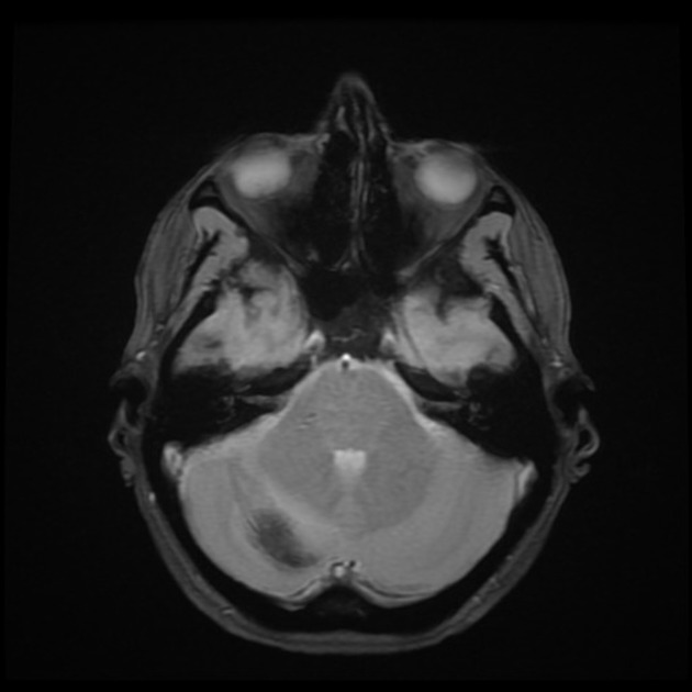
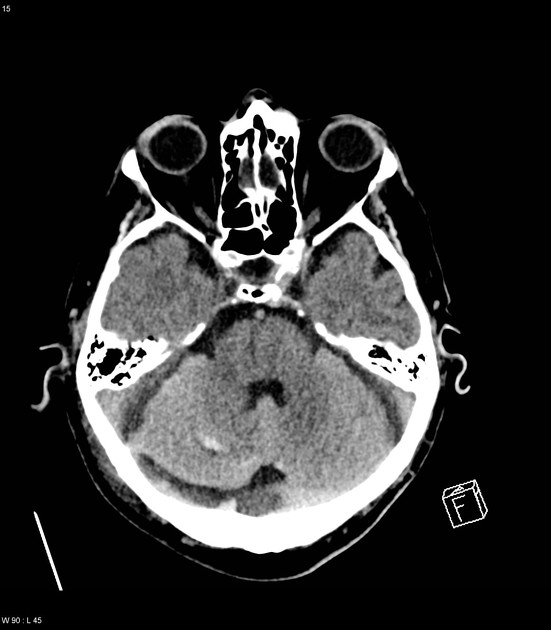
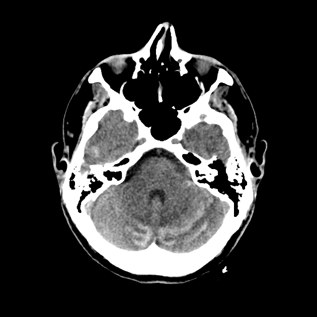
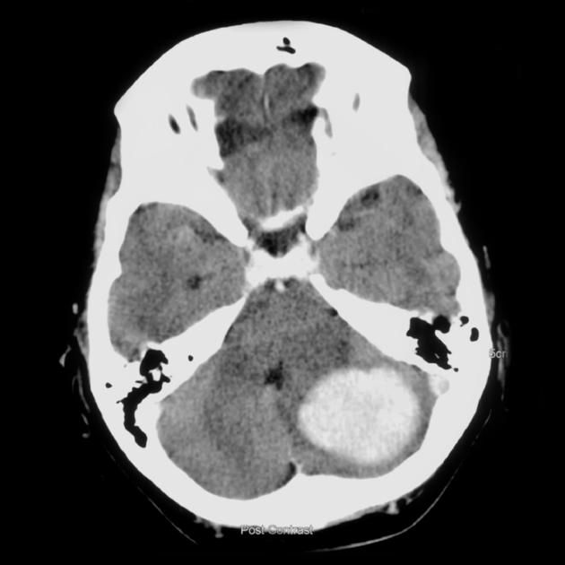
CT
The most common radiologic finding is layering of blood over superior folia, called the zebra sign 6,7, and less frequently it can be an intraparenchymal or lobar hemorrhage. Cerebellar hemorrhage can be contralateral or ipsilateral to the site of surgery, and less commonly can be bilateral or even can be isolated to the vermis. The right clinical context is invaluable for image interpretation.
MRI
The appearance of hemorrhage on MRI varies with time and to some degree the size of the hematoma (see aging blood on MRI).
Treatment and prognosis
Generally, no specific management is required 1. However, if the hemorrhage is large enough to cause obstructive hydrocephalus, then further neurosurgical intervention is required 1. It has a poor prognosis 8.


 Unable to process the form. Check for errors and try again.