
Presentation
ICU patient.
Patient Data
Age: 35 years
Gender: Male
From the case:
Pericardial fat tag sign
Download
Info
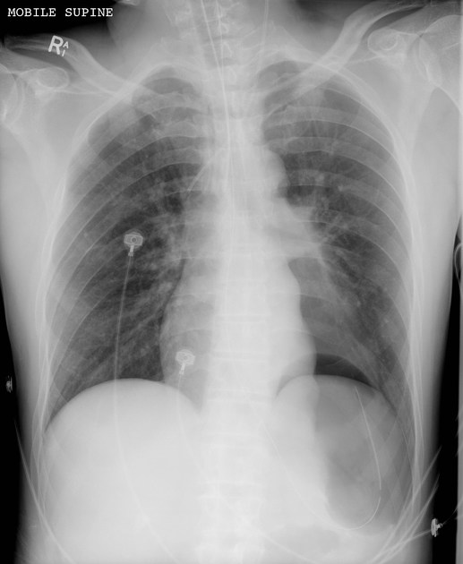
ETT tip is projected in the proximal right main bronchus. NGT and left jugular CVL are well positioned.
Left deep sulcus sign and pericardial fat tag sign indicating a pneumothorax. Allowing for patient rotation, no signs of mediastinal shift.
From the case:
Pericardial fat tag sign
Download
Info
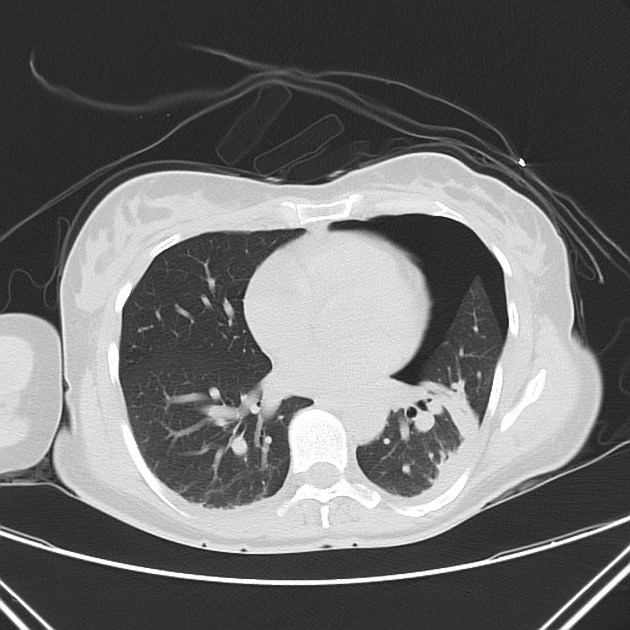
Pneumothorax confirmed on the left, with tags of pericardial fat at the cardiac apex hanging posteriorly. No mediastinal shift. Dependant LLL atelectasis.
Case Discussion
The pericardial fat pad sign is a nice but rare sign of pneumothorax on supine CXR. The pericardial fat displaces laterally as the lung is no longer adjacent to it pushing it against the mediastinum, hence causing a lumpy appearance of the cardiac borders.
Case courtesy of Dr Nivene Saad.


 Unable to process the form. Check for errors and try again.