
Presentation
Post CABG routine check CXR.
Patient Data
Age: 55 years
Gender: Male
From the case:
Retrodiaphragmatic pericardial fat tag sign
Download
Info
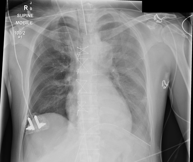
Patient is rotated.
Sternal wires and mediastinal clips in keeping with recent CABG. ETT, right jugular CVL, mediastinal drain and left basal ICC are well positioned.
Increased lucency at the right base, sharp contour of the right hemidiaphragm, deep sulcus sign and a right lower pericardial fat tag sign posterior to the medial aspect of the hemidiaphragm indicate a pneumothorax.
Increased density in the left hemithorax is due to a combination of patient rotation and left pleural effusion (known).
Case Discussion
The right pneumothorax was confirmed on CT and an ICC inserted.
Case courtesy of Dr Nivene Saad.


 Unable to process the form. Check for errors and try again.