
Presentation
Patient under treatment for chest infection for two months developed seizures with progressive deterioration of the level of consciousness.
Patient Data
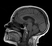

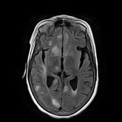
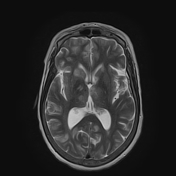
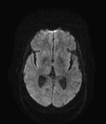
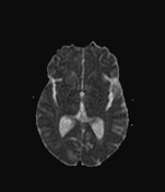
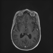
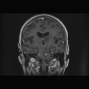
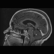
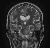

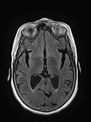
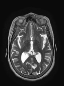
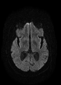
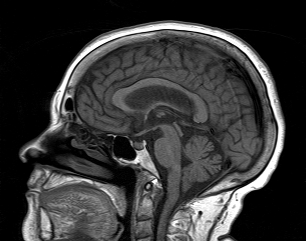
The MRI sequences demonstrate innumerable small intra-axial nodules at both infra- and supratentorial levels as well as brainstem and basal ganglia, ranging from 3 to 7 mm in diameter. They appear of intermediate to low signal on T1WI, high signal on T2WI, and FLAIR with low signal centrally, and surrounding edema. The postcontrast sequences show ring-like and nodular enhancement. Moderate dilatation of the ventricular system (3rd and lateral ventricles) with mild enlargement of the cerebral sulci.
Note a low position of the cerebellar tonsils indicating a Chiari I malformation (incidental finding).
Right otomastoiditis is also noted.

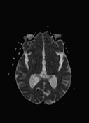
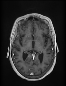
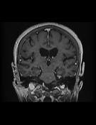
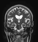
Significant improvement with decreased in size and number of the infra- and supratentorial lesions.
Case Discussion
Case of miliary cerebral tuberculosis most likely the result of the spread of primary pulmonary tuberculosis (miliary pulmonary tuberculosis proven in this case).


 Unable to process the form. Check for errors and try again.