Presentation
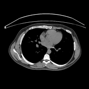
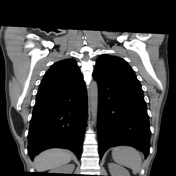
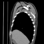
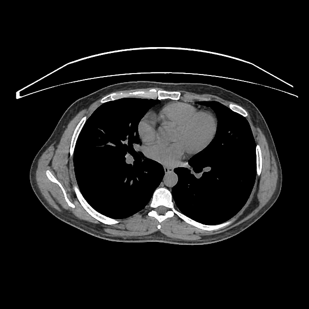
Progressive painless swelling of the right lateral chest wall.
Patient Data
Age: 70 years
From the case:
Intramuscular lipoma - latissimus dorsi


Download
Info

There is a well-circumscribed, homogeneous low attenuation mass (density= -110 HU) within latissimus dorsi muscle.
Case Discussion
CT features of a soft tissue lipoma.
The main differential diagnosis is a liposarcoma: usually with thick septa, enhancement or evidence of local invasion.


 Unable to process the form. Check for errors and try again.