
Patient Data
Age: Adult
Gender: Female
Note: This case has been tagged as "legacy" as it no longer meets image preparation and/or other case publication guidelines.
From the case:
Endometrioma, fibroid and ovarian cyst

Download
Info
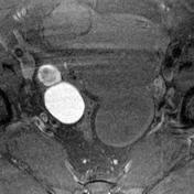
A well defined subserosal myometrial lesion, appearing isointense on T1W and hypointense on T2W images, is seen arising from right lateral wall of uterus, suggesting fibroid.
A well defined thin-walled cystic lesion is seen in right ovary, appearing hyperintense on T1 fat-suppressed sequence, and hypointense on T2W images ('shading sign'), suggesting endometrioma.
Left ovary shows a round lesion, hypointense on T1W and hyperintense on T2W images, suggesting simple ovarian cyst.
From the case:
Endometrioma, fibroid and ovarian cyst


Download
Info

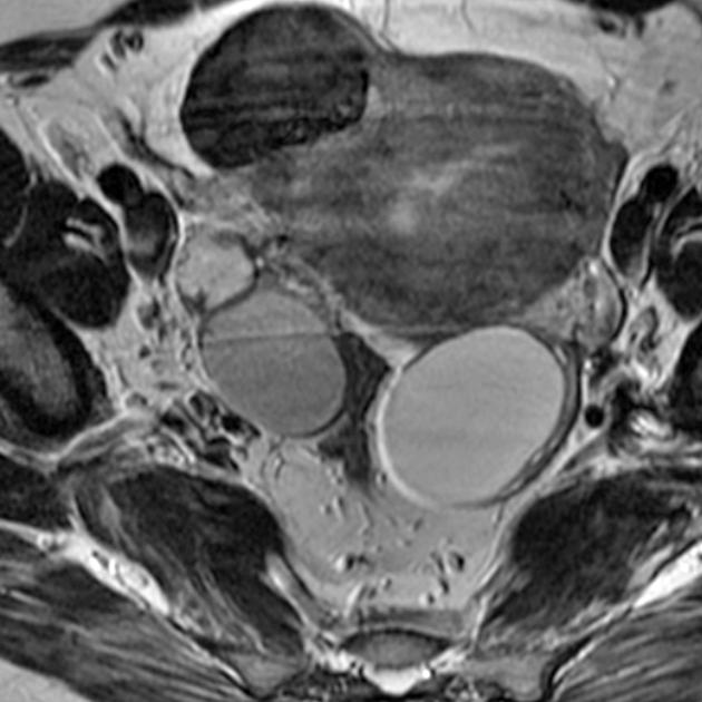
T2 annotated: E - endometrioma; F - uterine fibroid; C - ovarian cyst.
T1 FS annotated: E - endometrioma; F - uterine fibroid; C - ovarian cyst.


 Unable to process the form. Check for errors and try again.