
Presentation
Paraplagia and neck pain.
Patient Data
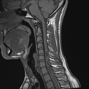

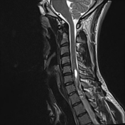
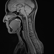

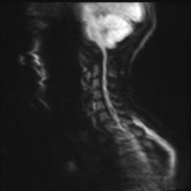
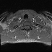
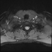

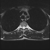
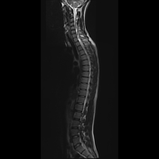

An eccentric dorsal cord-based mass lesion is noted with intramedullary and extramedullar components is seen opposite to C7/D1 level averaging roughly 19 x 9.5 mm, seen indenting cord from its posterior aspect. It is exerting T1 mild hypo and T2 iso intense signals. It indents manifestly the related cord with anterior displacement. The tumor nidus enhances avidly.
Cord syrinx is noted extending from C5 to D5 vertebral levels.
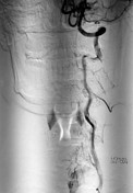


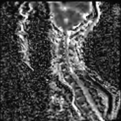
Digital subtraction angiography of the left vertebral artery shows a feeding vessel supplying the spinal mass originating from its proximal segment.

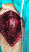
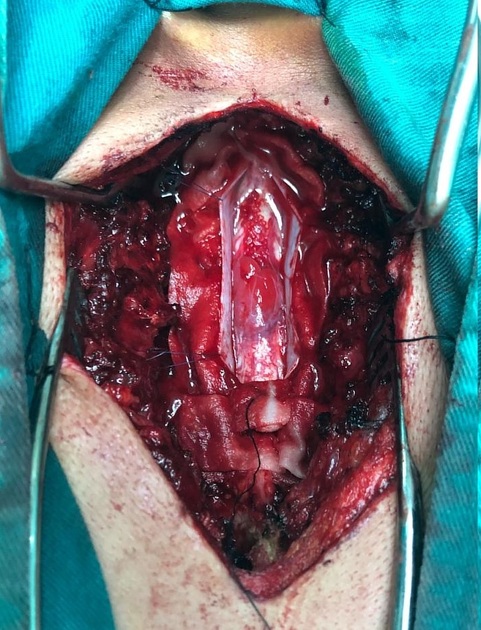
Operative photos showing a vascular spinal mass lesion at the cervical spinal cord.
Case Discussion
The imaging features shows a vascular spinal tumor with multiple tortious vascular channels within its substance and associated syrinx. The case was pathologically proved spinal hemangioblastoma - pathology report unavailable. The patient improved dramatically after the operation.
Case courtesy Dr. Mohamed Kaed, consultant radiologist, Dar Al-Ashaa, Alexandria, Egypt.
Case courtesy Dr. Mostafa Al-Askary, lecturer of neurosurgery, Alexandria university, Egypt.


 Unable to process the form. Check for errors and try again.