
From the case:
Neurofibromatosis type 1


Download
Info
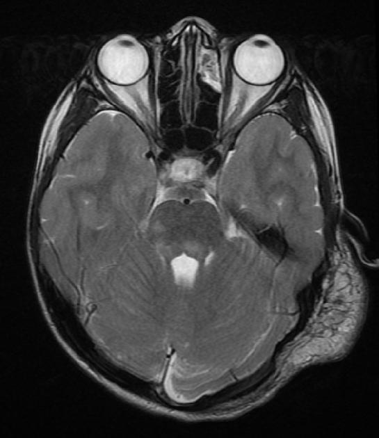
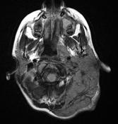
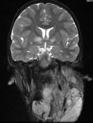
There is a plexiform neurofibroma involving the left side of the neck, scalp, and the submental/submandibular region. Note the multiple high T2 and FLAIR hyperintensity foci in the dentate nuclei, middle cerebellar peduncle, pons, midbrain, cerebral peduncles, and the basal ganglia.
Case Discussion
There is a plexiform neurofibroma (pathognomonic for NF1) involving the left side of the neck, scalp, and the submental/submandibular region. Note the multiple high T2 and FLAIR hyperintensity foci in the dentate nuclei, middle cerebellar peduncle, pons, midbrain, cerebral peduncles, and the basal ganglia.


 Unable to process the form. Check for errors and try again.