Presentation
Chronic low back pain radiating to right leg
Patient Data


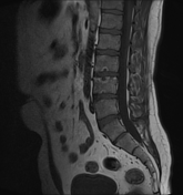
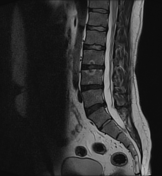
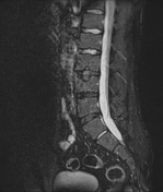
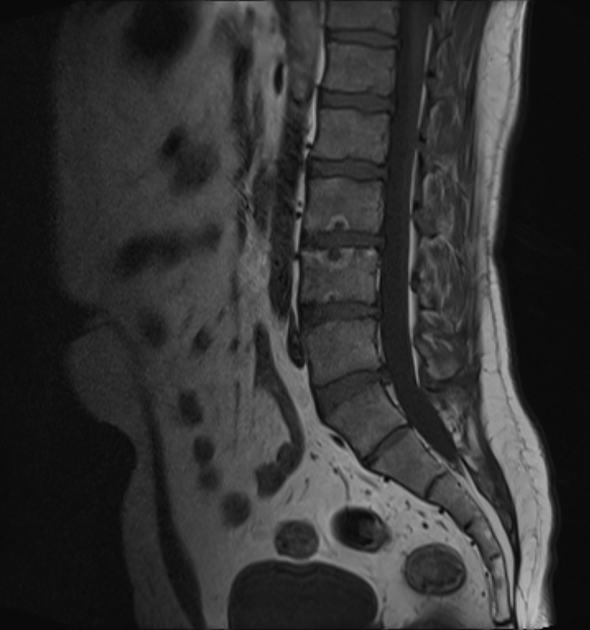
Endplate discontinuity at central part of inferior endplate of L3, superior endplate of L4 and inferior endplate of L4 vertebrae. These areas demonstrate peripheral T1 and T2 high signal with loss of signal on fat suppressed sequence (? fatty degeneration) and central T1 low and T2/STIR high signal and avid post contrast enhancement.
Mild disc dehydration at L3/4, L4/5 and L5/S1 levels.
Posterior disc bulge and small annular fissure (tear) at L5/S1 level resulting indentation on thecal sac and impression on right lateral recess.
Case Discussion
It has been suggested that the enhancement could be a result of continued prominent vascularity of granulation tissue within the disc fragment 1.


 Unable to process the form. Check for errors and try again.