
Presentation
Abdominal distension.
Patient Data
Age: 15 years
Gender: Male
From the case:
Peritoneal CSF pseudocyst


Download
Info

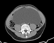
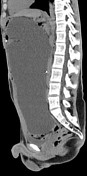
A large cyst fills the majority of the peritoneal cavity, displacing solid organs and bowel. A smaller cyst is along the inferior liver margin.
Two sets of VP shunt tubing are present: one follows a looped course along the liver dome, and the second terminates in the large peritoneal pseudocyst.
Case Discussion
Peritoneal CSF pseudocysts are a rare complication of VP shunts, and are comprised of a fibrous, non-epithelialized wall. Treatment involves surgical/percutaneous drainage or excision. Patients can present with abdominal symptoms related to mass effect/compression, infection, or shunt malfunction.


 Unable to process the form. Check for errors and try again.