
Presentation
Cough for 2 years with abnormal chest x-ray.
Patient Data


Mild mediastinal and hilar adenopathy.
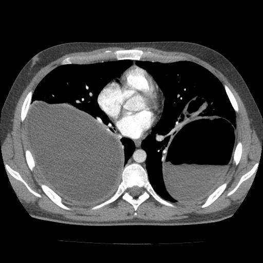
Right lower lobe thick-walled cyst measuring 13 x 15 x 16 cm; contains fluid only.
Left lower lobe thick-walled cyst measuring 16 x 10 x 14 cm; contains fluid, air and debris/floating membranes. There are a few adjacent inflammatory pulmonary opacities.
Trace pleural effusions.
Imaged abdomen is normal.
Case Discussion
Massive lower lobe hydatid cysts. The left lower lobe cyst contains air with a floating membrane, which indicates cyst rupture and communication with bronchioles. The right lower lobe cyst has not ruptured.
Hydatid disease in the lung is the second most common site (after liver). The lung can be infected by ingested oncospheres through a variety of mechanisms:
- lymphatic -> thoracic duct -> internal jugular vein -> right heart -> lungs
- portal vein -> sinusoids (if small) -> hepatic veins -> IVC -> right heart -> lungs
- transdiaphragmatic due to lymphatic spread or cyst rupture
- direct inhalation


 Unable to process the form. Check for errors and try again.