
Presentation
Bilateral leukocoria.
Patient Data
Age: 6 months
Gender: Male

Download
Info

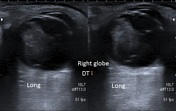
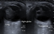
Right globe demonstrates a heterogenous structure filling the anterior segment of the viterous body with an echogenic band extending posteriorly to the optic disc. Arterial blood flow is seen within by color Doppler.
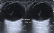
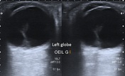
Left globe demonstrates an echogenic band in the posterior segment of the vitreous body in contact with the optic disc.
Case Discussion
The clinical presentation and ultrasound features are most consistent of bilateral persistent hyperplastic primary vitreous, more conspicuous on the right globe.


 Unable to process the form. Check for errors and try again.