
Presentation
Preoperative chest X-ray.
Patient Data
Age: 25 years
Gender: Female
Download
Info
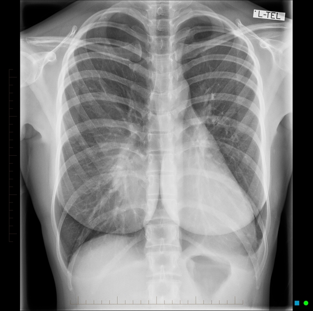
Classic features of pectus excavatum as demonstrated on this frontal chest X-ray.
Download
Info

Pectus excavatum is confirmed on this lateral view as demonstrated by the inward depression of the sternum.
Case Discussion
This case is an excellent example of the medial breast margin sign, which may be seen in women with pectus excavatum on frontal chest X-rays. This sign consists of more sharply defined and more vertically oriented medial breast borders compared to those of women with normal chest wall morphology.
This case also features the other classic signs of pectus excavatum, which include:
- increased density in the right inferomedial lung
- blurring of the right heart border
- vertical anterior ribs
- convex left heart border
- obliteration of the descending thoracic aortic silhouette


 Unable to process the form. Check for errors and try again.