
Presentation
Abnormal vaginal bleeding.
Patient Data


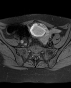

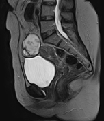
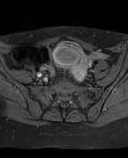
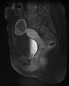
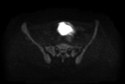
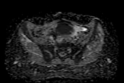
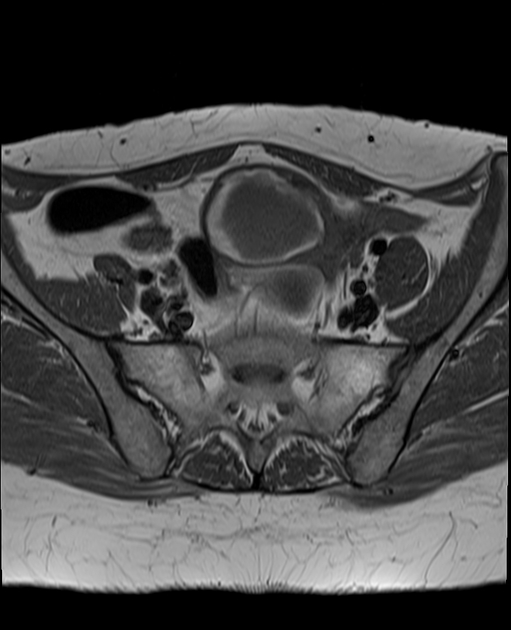
The MRI sequences demonstrate a well-defined suprauterine mass (5x4.5x4 cm) of intermediate signal on T1WI, heterogeneous high signal on T2WI with peripheral area of high signal on T1WI and low signal on T2WI (hemorrhagic area). Small adjacent cystic components are noted with hemosiderin sediment. The DWI shows a high signal with low ADC indicating restricted diffusion. No enhancement seen on postcontrast sequences. Minimal effusion is noted around the mass and in Douglas pouch.
Small intramural leiomyoma (8 mm) with no significant endometrial thickening seen.
The right ovary appears normal.
No pelvic lymphadenopathy is seen.
Case Discussion
The patient went on to have bilateral salpingo-oophorectomy with total hysterectomy. The histopathological exam with immunochemistry confirmed the diagnosis of adult granulosa cell tumor of the ovary.
Additional contributor: A.Ramdani, MD.


 Unable to process the form. Check for errors and try again.