
Presentation
50 years old man with end-stage renal failure, had inserted an internal jugular vein catheter on the left for dialysis.
Patient Data
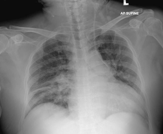
The left internal jugular vein catheter (IJC) was noted at the left neck and passing through the left mediastinum.
Consolidative changes at right mid-zone. No pleural effusion.
Thus CT thorax was performed to exclude IJC puncture to the aorta.
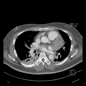

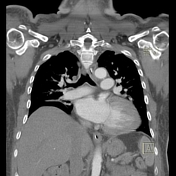
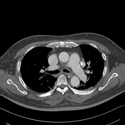
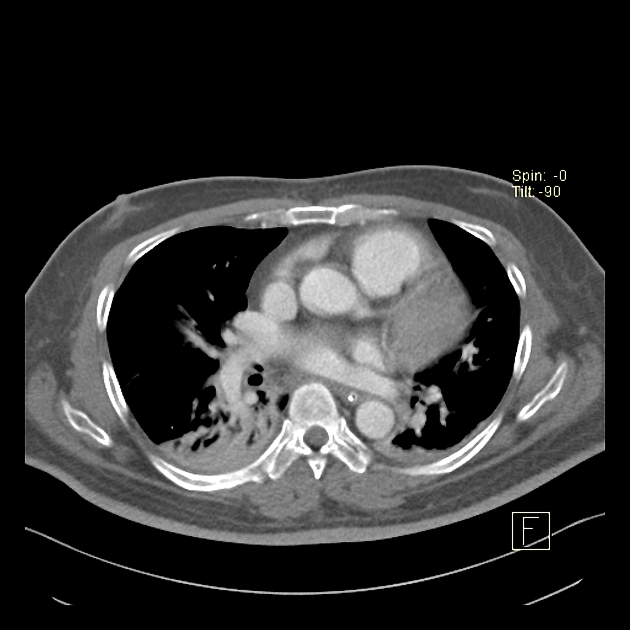
The left-sided catheter is seen coursing the left sternocleidomastoid pointing to posterior inferiorly. It enters the left subclavian vein then follow into the left brachiocephalic vein. The left superior pulmonary veins drain directly into the left brachiocephalic vein. The tip of the catheter is seen within this left superior pulmonary vein.
Case Discussion
Partial anomalous pulmonary venous return (PAPVR) is a rare condition in which some or partial pulmonary veins drain straight into the systemic circulation.
In this case, the left upper pulmonary veins are draining directly to the left brachiocephalic vein. Thus this created a vascular pathway that the internal jugular vein catheter (IJC) was going into.
The surgeon took the blood from the IJC and found it has high oxygenated blood. From the chest radiograph, one might mistake the IJC has punctured into the aorta, in which the IJC will give high oxygenated blood. Thus CT thorax with contrast was important for diagnosis. In this case, the high oxygenated blood comes from the pulmonary veins.


 Unable to process the form. Check for errors and try again.