
Presentation
Abdominal pain and distention.
Patient Data
Age: 75 years
Gender: Female
From the case:
Acute small bowel (ileal) volvulus


Download
Info

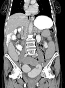
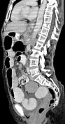
Diffuse dilation of small bowel with transition in the right mid abdomen with swirled appearance. Transition is at the distal/terminal ileum. 5 x 3 cm exophytic oval mass with heterogeneous enhancement and small fleck of calcification likely arising from the mid ileum in the left lower abdomen, a relatively short segment after it passes behind the volvulus. Vessels are swirled with the SMV wrapping around the SMA counterclockwise at the site of volvulus. Associated congestion in the mesentery with mild nodal enlargement. Small ascites.
Mild thickening/fluid associated with ascending colon.
Case Discussion
Small bowel volvulus resulting in acute obstruction.
Note:
- this patient is 75 years old - this is not just a problem in pediatrics!
- however, in this case, there is a 5 x 3 cm exophytic oval mass arising from the mid ileum (likely a GIST) which likely contributed to this patient developing the volvulus by tethering that segment of bowel.
- the location of volvulus is the distal ileum (remote from the mass)
- it is easiest to follow on the axial images
- the SMV wraps around the SMA counterclockwise, and thus has the opposite of normal relationship
- this results in vascular compression and mesenteric congestion
- the oral contrast gradually becomes more dilute in the distal small bowel


 Unable to process the form. Check for errors and try again.