
Presentation
Pelvic swelling with severe low back pain.
Patient Data


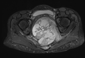
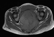
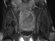
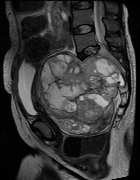
A huge well-defined heterogeneous solid lesion of mixed-signal intensity is demonstrated in the pelvis, arising from the sacrum. It shows heterogeneous postcontrast enhancement.
Case Discussion
Schwannomas are very rarely seen in the sacral region and constitute 1-5% of all spinal schwannomas. They are mostly benign lesions.
Giant invasive spinal schwannomas are masses that invade more than 2 vertebral levels, invade vertebral bodies and by extending posteriorly, reach the myofascial regions. They often present with pains and neurological symptoms.
MR findings, in this case, show a solid lesion of mixed-intensity with a heterogeneous contrast enhancement demonstrated anterior to the sacrum. It measures 14.0 x 13.1 x 9.8 cm. There is a foraminal extension of the mass with the destruction of the sacral vertebrae.


 Unable to process the form. Check for errors and try again.