
Presentation
Cough, lung mass on CXR.
Patient Data
Age: 20 years
Gender: Female
From the case:
Cystic pulmonary hydatidosis


Download
Info
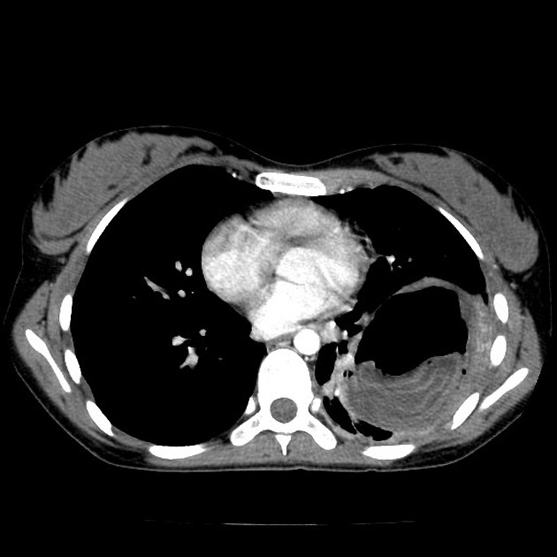
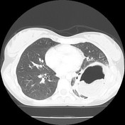
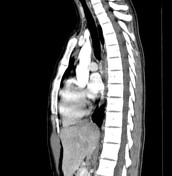
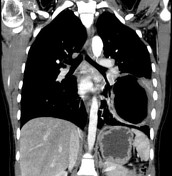
Large cystic mass in the left lower lobe with air-fluid level and decompressed dependent floating membrane. Mild mediastinal and left hilar adenopathy.
Case Discussion
Massive left lower lobe cystic mass containing a folded floating membrane, characteristic for hydatid cyst which has decompressed into the airway via a bronchiole (and thus contains air). Hydatid disease in the lung is the second most common site after liver.


 Unable to process the form. Check for errors and try again.