
Presentation
10 years of progressively growing mass covering the right orbit. Patient was born with a very dark birth mark in the temporal region.
Patient Data
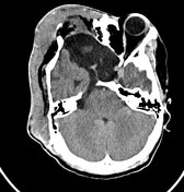

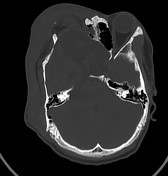
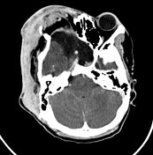
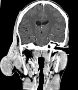
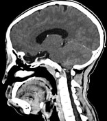
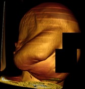
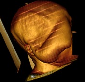
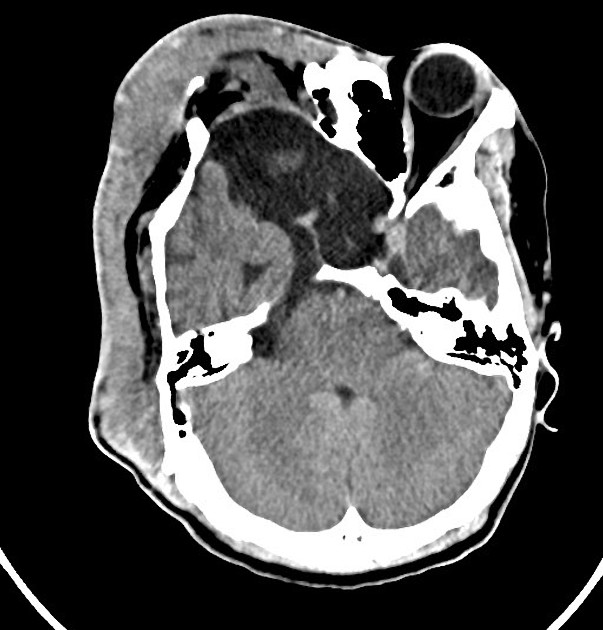
With respect to the very large facial mass, it involves the right orbit, face, and right cranial convexity resulting in thickened area of enhancement, and develops an exophytic component protruding along the rightward and lateral aspect of the face. There is a covering of the right globe which has dystrophic calcifications and abnormal shape indicating phthisis bulbi. There is some buckling and irregularity of the right optic nerve. There is loss of fat plane between the right masseter muscles and superficial parotid. The mass has very large veins along the posterior aspect draining into the right internal jugular vein, and several large arterial collaterals supplied by the external carotid artery.
Estimated size 13 x 4 x 14 cm. Loss of the normal osseous architecture of the sphenoid sinuses, right worse than left ethmoid sinuses, and right posterior lateral orbital walls with thinning/loss of bone. Hypoplasia of the right maxillary sinus. Deformity of the right zygoma. Thinning of the calvarium along the posterior right parietal region and somewhat diffusely along the right convexity. Thinning and expansion of the bone of the right orbit, with extension of the anterior temporal fossa into the typically expected location of the orbital cavity.
Case Discussion
Very large vascular mass involving the right orbit, face, and cranial convexity, with imaging features of a facial plexiform neurofibroma. This is compatible with the patient's history of slow growth and dark birthmark in the right temporal region (suggestive of cafe-au-lait macules, which are characteristic skin lesions of neurofibromatosis).


 Unable to process the form. Check for errors and try again.