
Presentation
Persistently rising prostate-specific antigen (PSA).
Patient Data


Multi-parametric magnetic resonance imaging mpMRI
Findings:
Quality: mild geometric distortion on DWI, does not compromise diagnostic confidence
Prostate size: 51 x 44 x 52 mm (CC x AP x ML) ≈61 mL.
Hemorrhage: none
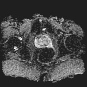
Peripheral zone (PZ): uniform hyperintense signal with one focal finding
Focal lesion #1:
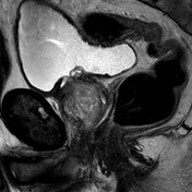
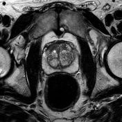
- location: right mid gland/apical anterior peripheral zone (PZa) and right anterior fibromuscular stroma (AFMS), visible on axial images (ima 11-14) and sagittal images (ima 17)
- lesion size: 17 x 9 x 13 mm
- T2w: circumscribed, homogenous moderate hypointense focus - category 5/5
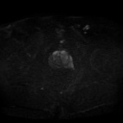
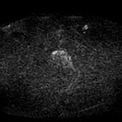
- DWI: markedly hyperintense on DWI (b1000 and b1400) and markedly hypointense on ADC focus - category 5/5
- DCE: focal early enhancement - positive
Transition zone (TZ): moderate heterogeneity, multiple BPH nodules with one focal finding
Focal lesion #2:
- location: mid basal anterior fibromuscular stroma (AFMS), visible on axial images (ima 17-18), sagittal images (ima 13-15)
- lesion size: 13 x 8 x 16 mm
- T2w: circumscribed, homogenous moderate hypointense focus - category 5/5
- DWI: markedly hyperintense on DWI (b1000 and b1400) and markedly hypointense on ADC focus - category 5/5
- DCE: focal early enhancement - positive
Prostate margin: lesion #1 with broad capsular contact (~16 mm)
Overall PI-RADS category: 5
Neurovascular bundles: not involved
Seminal vesicles: not involved
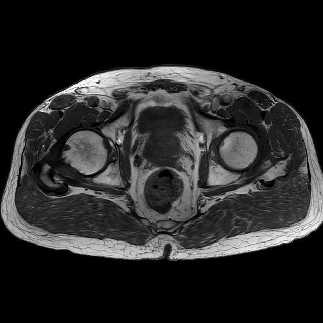
Lymph nodes: no enlarged or suspicious lymph nodes
Pelvic bones: typical “herniation pit” at the anterior aspect of the superolateral femoral head-neck junction, subchondral cyst of the symphysis pubis, no signs of bony metastasis
Impression:
Very highly suspicious lesion of the right apical, anterior peripheral zone (PZa) with broad capsular contact and further lesion of the mid basal anterior fibromuscular stroma (AFMS) - PI-RADS 5.
A prostate biopsy was recommended.

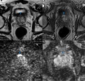
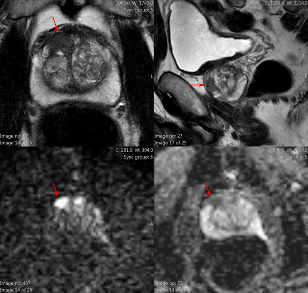
Key images:
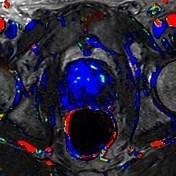
Focal lesion #1 (red arrows):
- circumscribed, homogenous moderate hypointense focus, markedly hyperintense in high b-value (b1400) and markedly hypointense in ADC (red arrows) with early enhancement on DCE (not shown),
- located in the right mid gland/apical anterior peripheral zone and right anterior fibromuscular stroma (AFMS)
Focal lesion #2 (blue arrowheads):
- circumscribed, homogenous moderate hypointense focus, markedly hyperintense in high b-value (b1400) and markedly hypointense in ADC (blue arrowheads) with early enhancement on DCE,
- located in the middle (bilateral) of the basal anterior fibromuscular stroma (AFMS)
Case Discussion
This case demonstrates a PI-RADS 5 lesion of the right apical anterior peripheral zone (PZa) and a further PI-RADS 5 lesion of the basal anterior fibromuscular stroma (AFMS).
Histology of the MR in-bore revealed a continuous infiltrate of an acinar adenocarcinoma (modified Gleason score 4+3=7b, G2b, high grade) within the two cores.
After consultation with his urologist patient is receiving radiation therapy.


 Unable to process the form. Check for errors and try again.