
Presentation
Progressive painless swelling of the medial aspect of the left leg.
Patient Data
Age: 45 years
Gender: Male
From the case:
Dermatofibrosarcoma protuberans (DFSP)


Download
Info

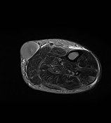
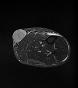
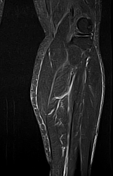
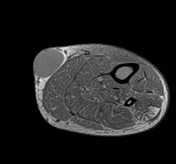
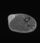
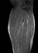
The MRI sequences demonstrate a well-defined subcutaneous ovoid mass of the medial aspect of the left leg (4.5x4.2x2.8 cm). It shows an intermediate signal on T1WI, high signal on T2/STIR with intense and heterogeneous enhancement on postcontrast sequences. No involvement of the adjacent medial gastrocnemius muscle.
Case Discussion
MRI features of a subcutaneous tumor, pathologically proven as a dermatofibrosarcoma protuberans (DFSP)


 Unable to process the form. Check for errors and try again.