
Cerebral arteriovenous malformation at posterior body of corpus callosum
Presentation
Headache, vomiting
Patient Data
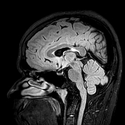
MRI + MRA Brain


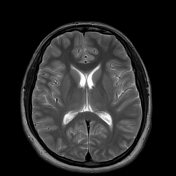
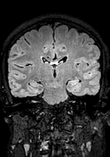
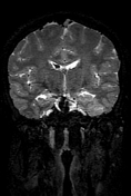
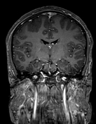
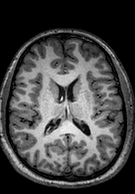
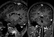
Wedge-shaped clump of variable hyperintense signal is noted in anterior body of right lateral ventricle.
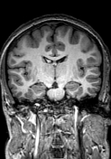
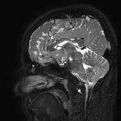
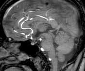
Serpiginous collection flow voids is identified along upper margin of posterior body of corpus callosum, producing bag of worms appearance, covering an area measuring 2 x 1.5 cm. Posterior body of corpus callosum is comparatively thinner than its other parts.
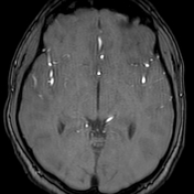
After administration of I/V contrast—
No abnormal enhancement is noted in brain parenchyma.
Contrast material is identified inside right lateral ventricle.
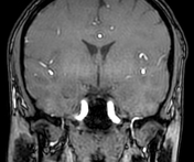
Feeding artery to the bag of worms is identified arising from A3 segment of right anterior cerebral artery (ACA), and draining vein is identified as posterior callosal vein. The bag of worms also communicates with intraventricular veins through perforating veins traversing across posterior body of corpus callosum.
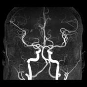
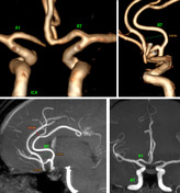

A2 segment of left ACA artery courses almost 6-12 mm anterior to right ACA instead of being parallel.
Also, proximal 1 cm of left A2 segment lies to the right of midline, flipping anterior & overlapping its right counterpart.
Anterior communicating artery (AComA) is located near the end of A2 segment, instead of at its root.
Case Discussion
Differential Dx : Arteriovenous fistula (AVF)
Feeder of AVM : A3 segment of right anterior cerebral artery (ACA)
Drainage : Posterior callosal vein + Subependymal veins of right lateral ventricle
Additional Points
Leakage of I/V contrast into right lateral ventricle indicates intraventricular hemorrhage, most likely arising from the abnormal perforating vessels connecting posterior callosal vein with subependymal veins.
It is likely that there is pressure asymmetry between right & left ACA arteries, due to which none of their segments are parallel to each other. The pressure difference forces the left A2 segment to flip over its right counterpart and lie to the right of the midline! Also, it runs clearly about 1 cm anterior to its counterpart.


 Unable to process the form. Check for errors and try again.