
Presentation
Presented with a complaint of the left little finger nodule on the palmar side for the last months. No trauma/ pain/ skin changes/ tingling.
Patient Data
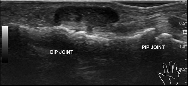
There is a well-defined lobulated lesion in the region of interest.
Size
- 14 mm (length) x 10 mm (transverse) x 5 mm (anteroposterior)
Location
- the lesion is on the palmar side of the distal interphalangeal joint level
- it shows mild dorsal side extension on the ulnar side
- it abutts the flexure tendon
- ulnar side neuro-vascular bundle is elevated by the lesion
- it is separate from tendon/bone/joints
Dynamic scan
- free flexure tendon movements from the lesion
Morphology
- solid
- hypoechoic, mild heterogeneous
- posterior acoustic enhancement present
- no calcification / cystic changes
- not compressible
Doppler
- no intralesional flow signals
- both digital arteries are patent
Adjacent bone cortex
- no erosion
Flexure tendon
- intact with normal echopattern
- no tenosynovitis
Interphalangeal joints
- no effusion
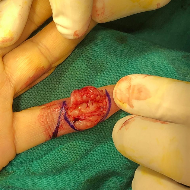
There is a lesion on the palmar side of the little finger. It overlies the middle phalanx region. The gross specimen photos show complete excision.
Case Discussion
An adult female presented with a painless finger nodule. Ultrasound features favored a possibility tenosynovial giant cell tumor. Surgical excision of the lesion was done and histopathology confirmed the lesion being tenosynovial giant cell tumor. It is the most common soft-tissue lesion of the hand and wrist 1.
Intraoperative and gross pathology photos courtesy: Operating surgeon Dr. Nisarg A. Patel.


 Unable to process the form. Check for errors and try again.