
Presentation
History of horizontal nystagmus on the right. Ophthalmologic evaluation revealed hypoplasia of the right optic nerve.
Patient Data

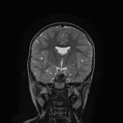
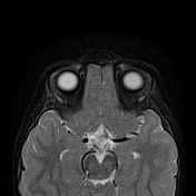
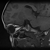

Hypoplasia of the right optic nerve (including the posterior part of the intraorbital segment, intracanalicular and intracranial segments) and optic chiasm (best evaluated on coronal T2-weighted images).
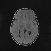
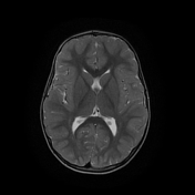
Lack of a septum pellucidum with concomitant mild flattening of the anterior horns of the lateral ventricles and a downward-sloping lower aspect of the anterior part of the horns (best evaluated on coronal T2-weighted images).
Case Discussion
This case demonstrates typical features of septo-optic dysplasia: hypoplasia of the right optic nerve and small optic chiasm, and absent septum pellucidum with resultant typical configuration of the anterior horns of the lateral ventricles.


 Unable to process the form. Check for errors and try again.