
Presentation
Chronic headaches and left hearing loss.
Patient Data
Age: 70 years
Gender: Female
From the case:
Meningioma of the cerebellopontine angle


Download
Info

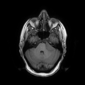
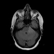
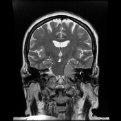
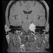
There is a well-defined extra-axial dural-based mass of the left cerebellopontine angle, enlarging the porus acusticus with extension into the internal auditory canal. It displays an isosignal to the cortical grey matter on T1WI / T2WI with an avid homogeneous enhancement on postcontrast sequences with a dural tail sign. A mass effect is noted on the brainstem and middle cerebellar peduncle as well as the VII / VIII nerve roots.
Axial T1 post-contrast images also demonstrate herringbone artefact.
Case Discussion
MRI features of a meningioma of the left cerebellopontine angle extending into the ipsilateral internal auditory canal.


 Unable to process the form. Check for errors and try again.