
Presentation
Chronic cough and history of chronic sinusitis.
Patient Data
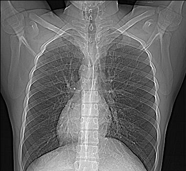
CT scout imaging demonstrates:
- right-sided heart with linear shadows seen at both lung fields (more evident at left paracardiac region) suggestive of bronchiectasis
- relatively hyperinflated lungs


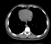
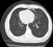
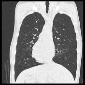
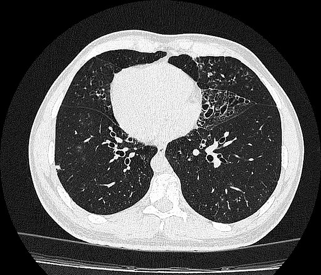
CT reveals situs inversus with dextrocardia. There is also cystic bronchiectasis in the middle and anterior zones of both lungs.
Also, the centrilobular ground-glass opacities and the tree-in-bud sign are well visible in both left middle and lower lobes, as well as right ligula segments and lower lobe, suggests current endobronchial infection.
Pleural based ground-glass nodules are seen in the left upper lobe anterior segment and the right lower lobe superior segment.

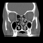
Frontal sinuses are not pneumatized.
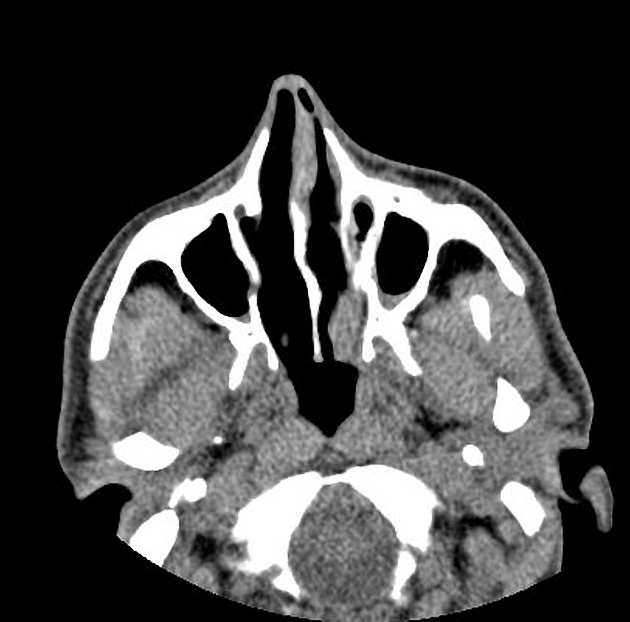
Mild mucosal thickening is present at maxillary and sphenoidal sinuses.
Nasal septal deviation to the right side with the left-sided spur formation is noted.
Case Discussion
A typical case of Kartagener syndrome, including chest and paranasal sinus CT scans.
The triad of situs inversus, bronchiectasis and sinusitis are clearly shown in this case.


 Unable to process the form. Check for errors and try again.