
Presentation
Low-risk prostate cancer detected transurethral resection of the prostate (TURP). Radiation therapy planning.
Patient Data


Findings:
Quality: no major artifacts
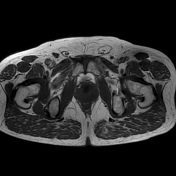
Prostate size: 44 x 44 x 54 mm (CC x AP x ML) ~54 mL.
Hemorrhage: none
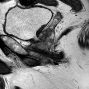
Peripheral zone (PZ):
- two in T2 hypointense foci, hyperintense in b1400, hypointense in ADC
- right apical and midglandular anterior and posterolateral zones
- lesion size: ~25 x 15 x 15 and ~9 mm
Transition zone (TZ):
- central periurethral defect after transurethral resection of the prostate (TURP)
- some BPH nodules bilaterally in the midglandular and basal posterior transition zone (TZa)
- indistinct margin of the pseudocapsule in the right apical and midglandular transition zone
Prostate margin: broad capsular contact, no bulge or breach
Neurovascular bundles: symmetric
Seminal vesicles: no signs of invasion
Lymph nodes: oval external iliac lymph nodes (up to 6 mm size, smooth margins)
Impression:
Suspicious focus in the right apical anterior and posterolateral as well as a second, smaller focus in the midglandular posterolateral peripheral zone (PZpl), consistent with the known prostate cancer.
An extraprostatic extension is possible.
MRI putative stage: cT3aNxMx

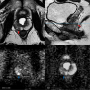
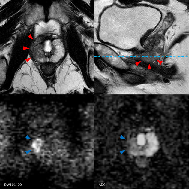
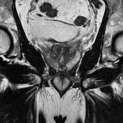
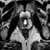
Key findings:
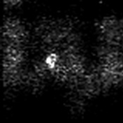
- small focus in the right midglandular posterolateral zone (arrows)
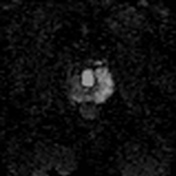
- large focus in the right apical (and midglandular) anterior and posterolateral zones (arrowheads)
- both lesions are hyperintense in b1400 and hypointense on ADC (blue arrows and arrowheads)
Case Discussion
This case illustrates a proven prostate cancer, accidentally detected on transurethral resection of the prostate (TURP).
Histology showed an acinar adenocarcinoma (modified Gleason score 3+3=6, G2a low grade ).
MRI was done for radiation therapy planning and staging purposes and shows two foci in the right apical and midglandular anterior and posterolateral peripheral zones.
Additional note:
Do not be fooled by Gleason score 3+3=6 revealed by the histology of the transurethral resection of the prostate (TURP).
This is a fairly large lesion and should be considered as a clinically significant cancer 1,2.
There is a broad capsular contact of the large lesion with possible extraprostatic extension.
They got the histology only from the tip of the iceberg.
The patient receives intensity-modulated radiotherapy.


 Unable to process the form. Check for errors and try again.