
Presentation
Ultrasound was requested for assessment of pyelonephritis. Normal renal function tests.
Patient Data
Both kidneys are of normal site, size and shape. They show increased parenchymal echo-pattern with decreased cortico-medullary differentiation. No stones or masses. No pelvicalyceal dilatation bilaterally.
The right kidney averages 6.5 x 2.6 cm (Length x TS) with a parenchymal thickness of 1 cm.
The left kidney averages 7 x 3.2 cm (Length x TS) with a parenchymal thickness of 1.2 cm.
Diffuse bilateral renal tiny parenchymal cysts, largest averages 3 mm with increased parenchymal echopattern, suggestive of autosomal recessive polycystic kidney disease.
Minimal bilateral perirenal collections, related to the lower pole of both kidneys, more on the right side, that raise the possibility of associated bilateral renal pyelonephritis.
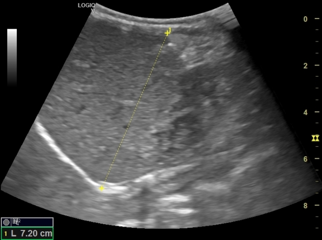
The liver is average in size (7.2 cm in MCL) with mild fibrotic changes. The spleen is of average size. The gallbladder is normal.
Case Discussion
Ultrasound demonstrates typical imaging features of autosomal recessive polycystic kidney disease. The kidneys appear echogenic likely due to the posterior acoustic enhancement of the tiny interstitial cystic changes with preserved reniform shape. The liver shows mild fibrotic changes. Bilateral mild perirenal fluid collections raise the possibility of associated pyelonephritis.


 Unable to process the form. Check for errors and try again.