
Presentation
Abdominal pain
Patient Data
Age: 50 years
Gender: Male
From the case:
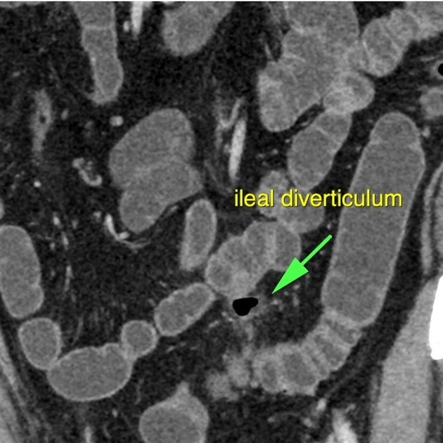
Ileal diverticulitis

Download
Info

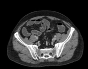
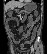
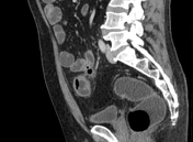
In the midline of the lower abdomen is a focus of central gas content and perifocal fat stranding adjacent an ileal loop mesenteric border with a tiny gas-filled connection to bowel lumen. Mild reactive thickening of the adjacent bowel wall. No abscess formation. No other diverticula.
Multiple gallstones.
Grade I lytic spondylolisthesis of L4 over L5.
Case Discussion
Imaging features of ileal diverticulitis is similar to colonic diverticulitis. Acute diverticulitis of the small bowel is rare. The patient was managed conservatively.


 Unable to process the form. Check for errors and try again.