Presentation
Right sided ptosis, ophthalmoplegia and decreased right eye movement
Patient Data
![](https://prod-images-static.radiopaedia.org/images/52253915/3003f8eb52d069696a5091b907b048_thumb.jpeg)
![This study is a stack](/packs/stack-YQKLCKBI.gif)
![](https://prod-images-static.radiopaedia.org/images/52253930/13c225139b5404b317463e95e3634f_thumb.jpeg)
![This study is a stack](/packs/stack-YQKLCKBI.gif)
![](https://prod-images-static.radiopaedia.org/images/52253950/f8cb2d9213a9aa2afa919f8e7a3525_thumb.jpeg)
![This study is a stack](/packs/stack-YQKLCKBI.gif)
![](https://prod-images-static.radiopaedia.org/images/52253968/b6ed6b1dfce86e502c38cd6e52195b_thumb.jpeg)
![This study is a stack](/packs/stack-YQKLCKBI.gif)
![](https://prod-images-static.radiopaedia.org/images/52253984/eae9296b5ceca4b1b306d335750c10_thumb.jpeg)
![This study is a stack](/packs/stack-YQKLCKBI.gif)
![](https://prod-images-static.radiopaedia.org/images/52254067/049aae1f314b3bd39c56b8158b19fa_thumb.jpeg)
![This study is a stack](/packs/stack-YQKLCKBI.gif)
![](https://prod-images-static.radiopaedia.org/images/52254231/4a5fd5b1d1d8a77fbce4e17fa2443f_thumb.jpeg)
![This study is a stack](/packs/stack-YQKLCKBI.gif)
![](https://prod-images-static.radiopaedia.org/images/52254332/e68d04d1b8d60b82886a833cb62173_thumb.jpeg)
![This study is a stack](/packs/stack-YQKLCKBI.gif)
![](https://prod-images-static.radiopaedia.org/images/52254354/09a09ff04a84b5180983ecbe325c05_thumb.jpeg)
![This study is a stack](/packs/stack-YQKLCKBI.gif)
![](https://prod-images-static.radiopaedia.org/images/52253915/3003f8eb52d069696a5091b907b048_big_gallery.jpeg)
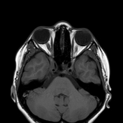
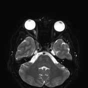
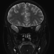
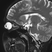
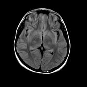
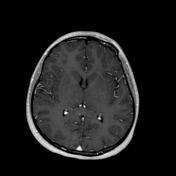
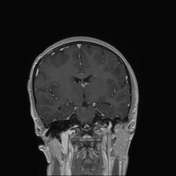
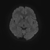
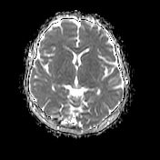
There is an intensely enhancing thickening in the right cavernous sinus region slightly extending to the right superior orbital fissure and right orbital apex. It causes enlargement of the right cavernous sinus and narrowing of the cavernous part of the right internal carotid artery. The mentioned thickening appears isointense to muscles on T1 and T2.
No cavernous sinus, superior ophthalmic vein or dural venous sinuses thrombosis. No aneurysm or CC fistula. No masses. No frank proptosis.
Normal looking pituitary gland, left cavernous sinus and suprasellar region.
Case Discussion
Appearances are those of Tolosa-Hunt syndrome.
Other differentials like meningioma, sarcoidosis, lymphoma, TB meningitis and pituitary gland tumor are unlikely in this case.