
Presentation
Shortness of breath and cough. Smoker. Pneumothorax?
Patient Data
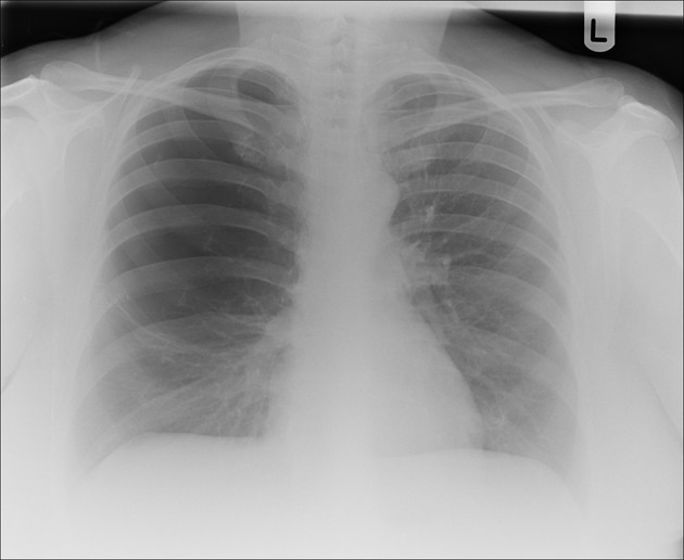
Large radiolucency area in the right upper and mid zones. Increased density of bronchovascular markings in the right lower zone.
Heart size normal. Left lung clear.
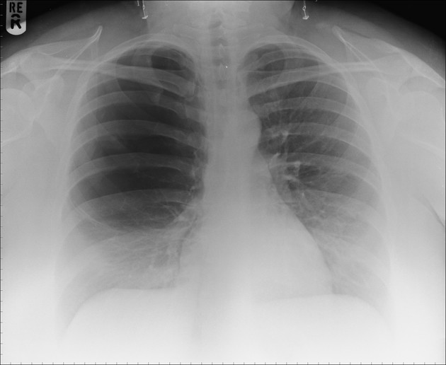
The appearances are essentially identical 10 years previously.
Case Discussion
Large isolated right upper lobe bulla. The left lung is entirely normal in appearance.
In the acute setting for the unprepared, this may be mistaken for a pneumothorax and lead to pleural drain insertion.
Two ways at least to avoid this mistake:
1. Take a look for old films. It might sound boring and repetitive, but it is so helpful, so often.
2. In this case, the increased bronchovascular markings in the lower zone are the normal lobes compressed. In a pneumothorax of this size the lung normal collapses towards the hilum.
This kind of bullous disease would be a candidate for bullectomy.


 Unable to process the form. Check for errors and try again.