
Presentation
Pain in the oral cavity associated with an indurated mucosa of the right retromolar trigone
Patient Data
Age: 70 years
Gender: Male
From the case:
Retromolar trigone squamous cell carcinoma


Download
Info
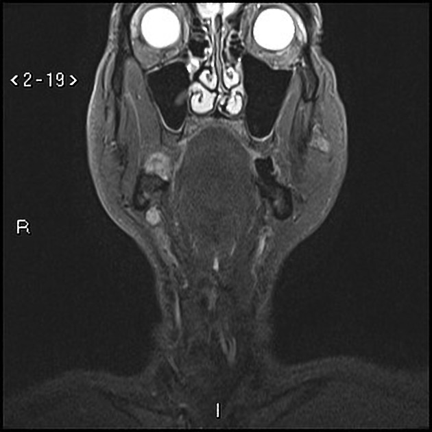
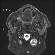
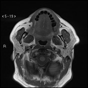
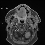
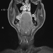
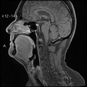
There is a rather well-defined small lesion within the right retromolar trigone, measuring 19 x 16 x 14 mm in its maximum dimensions. The lesion elicits a low signal on T1WI and a high signal on T2WI with mild post-contrast enhancement. There is no evidence of invasion of the surrounding structures.
There is no cervical lymphadenopathy.
Case Discussion
Pathologically proven right retromolar region SCC (T1 N0).


 Unable to process the form. Check for errors and try again.