
Presentation
Right shoulder lump.
Patient Data
Age: Adult
From the case:
Lipoma - subdeltoid

Download
Info
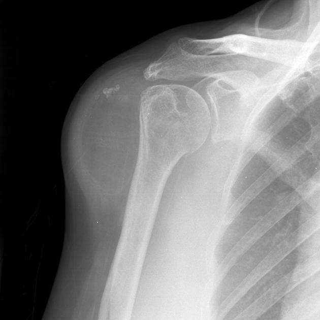
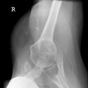
Xrays of the shoulder demonstrate a low attenuation (lower than muscle, similar to fat) deep to the deltoid muscle. At the superior margin of the lesion is an area of dystrophic calcification. No bony erosion.
From the case:
Lipoma - subdeltoid

Download
Info

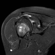
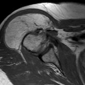
MRI of the shoulder (selected images only) confirms the mass to be predominantly of tissue returning fat-signal with suppression on STIR. Only thin whispy vascular enhancement is seen. No solid enhancing/non-fatty component.
Case Discussion
These features are characteristic of a lipoma or less likely low-grade liposarcoma located deep to the deltoid muscle.
Follow-up over a number of years demonstrated no growth or local symptoms, making the diagnosis of a lipoma.


 Unable to process the form. Check for errors and try again.