
Presentation
Chronic dry cough and chest discomfort. No hemoptysis, dysphagia, fever or weight loss.
Patient Data
Age: 20 years
Gender: Female
From the case:
Bronchogenic cyst

Download
Info
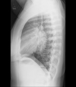
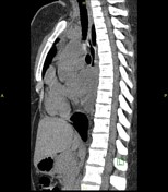
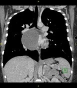
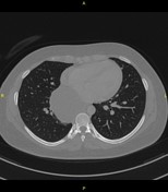
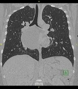
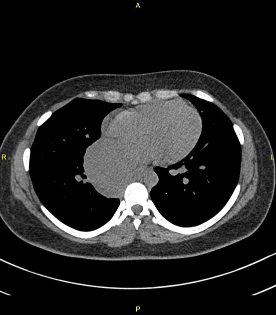
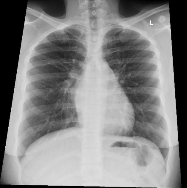
Large well-defined homogeneous non-calcified subcarinal middle and posterior mediastinal soft tissue density mass. Splaying of the carina. Clear lungs.
From the case:
Bronchogenic cyst

Download
Info
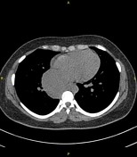
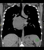
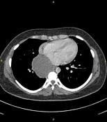
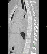
- Large well-defined extra-pulmonary subcarinal non-enhancing cystic mass having an average density of 28 HU (likely due to the proteinaceous contents).
- Non–calcified
- Thin imperceptible walls
- Splaying of the carina and mass effect over the main bronchus.
-
These radiological features are suggestive of a bronchogenic cyst.
Imaging differential diagnoses include esophageal duplication cyst, and pericardial cyst.
Download
Info
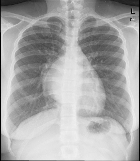
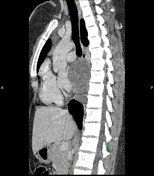
Subcarinal lesion seen on previous chest radiograph and CT chest is no longer appreciable.
Case Discussion
The lesion was resected.
Histopathology: Cyst lined by ciliated respiratory-type epithelium with subepithelial seromucous glands and smooth muscle, consistent with a bronchogenic cyst.


 Unable to process the form. Check for errors and try again.