
Patient Data
Age: 60 years
Gender: Male
Note: This case has been tagged as "legacy" as it no longer meets image preparation and/or other case publication guidelines.

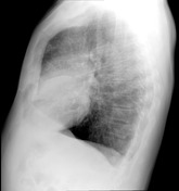
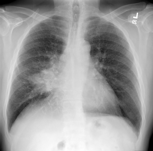
From the case:
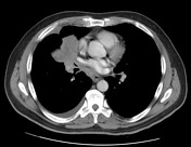
Lung cancer - adenocarcinoma

Download
Info
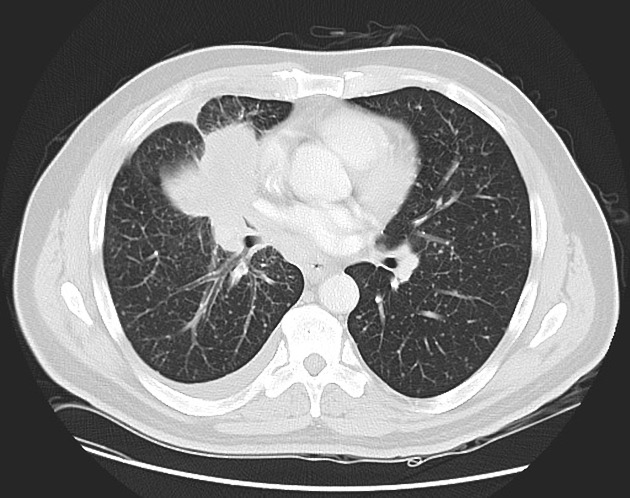
Selected images from a CT of the chest confirm a large hilar mass, consistent with a primary malignancy.
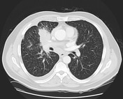
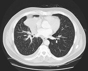
From the case:
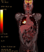
Lung cancer - adenocarcinoma

Download
Info
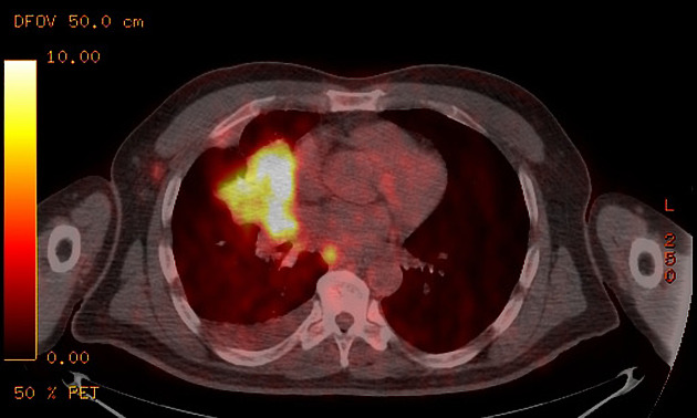
CT PET confirms increased metabolic activity in the mass as well as a subcarinal node.
Case Discussion
The patient went on to have a biopsy which pathologically proved an adenocarcinoma of the lung.


 Unable to process the form. Check for errors and try again.