
Patient Data
Age: Young adult


Download
Info
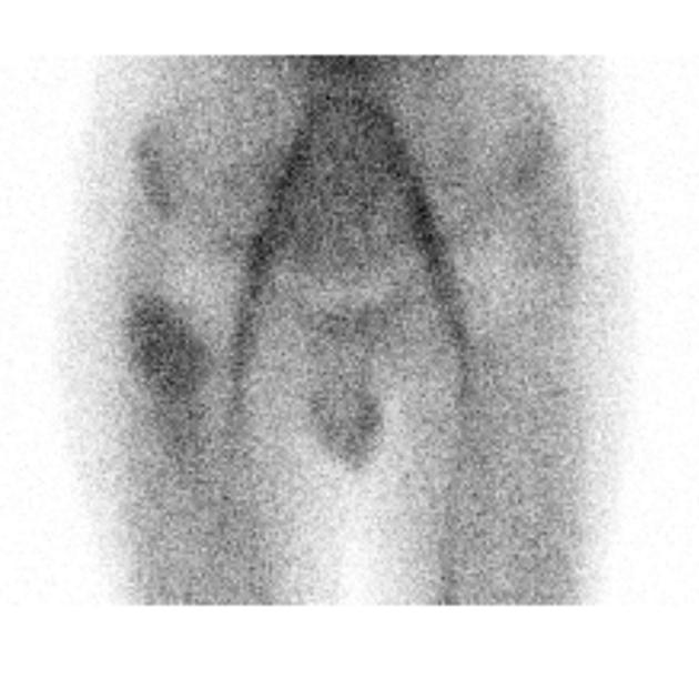
Bone scan (pool and delayed) images demonstrate increased uptake in the proximal right femur.
From the case:
Ewing sarcoma - proximal femur

Download
Info

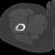
Scout and CT demonstrate a lytic agressive process involving the proximal femur.
From the case:
Ewing sarcoma - proximal femur


Download
Info
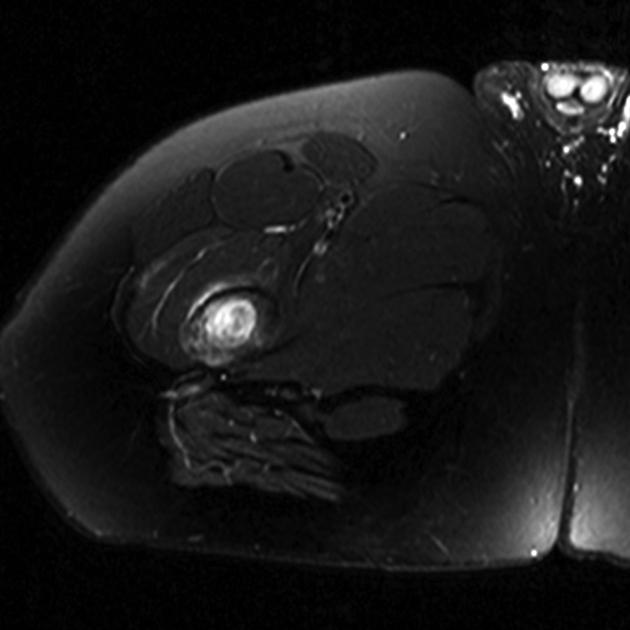
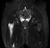
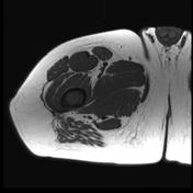
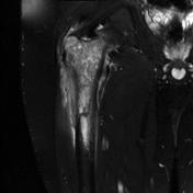
MRI confirms the CT findings and also demonstrates an extra-osseous component.
- Note: This case has been tagged as "legacy" as it no longer meets image preparation and/or other case publication guidelines.
Case Discussion
This case was confirmed to be of a Ewing sarcoma in a young adult.


 Unable to process the form. Check for errors and try again.